The central course of the nervus octavus and its influence on motility / by C. Winkler.
- Cornelis Winkler
- Date:
- 1907
Licence: In copyright
Credit: The central course of the nervus octavus and its influence on motility / by C. Winkler. Source: Wellcome Collection.
Provider: This material has been provided by King’s College London. The original may be consulted at King’s College London.
189/256 page 185
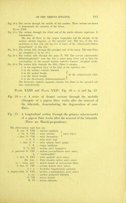
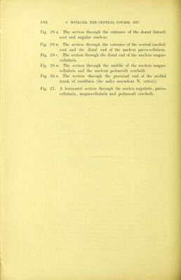
![of the contra-lateral fillet. Fig. 28 b. The section through the middle of this nucleus. Those sections are drawn to demonstrate the extensity of the lesion. Plate XXII. Fig. 28 c. The section tluough the distal end of the nuclei olivares superiores. It demonstrates: The loss of fibres in the corpus trapezoides and the atroph}' of the nucleus olivaris superior, at the operated side. The loss of the stria medullaris at that side and the loss of fibres of the //aberrirende Seiten- strangbiindel at that side: Fig. 28 D. The section falls through the proximal end of the lesion The same fibres are atrophied as in fig. 28 c. Fig. 28 E. The section falls through the genu N. VII. The crossed //aberrirendes Seitenstrangbundel now has lost a great many filjres and so have the surroundings of the crossed nucleus ventralis lemnisci (atrophied itself). Fig. 28 F. The section falls through the fillet. There is atrophy. 1 in the superficial layer of the fillet at the operated side. 2 in the nucleus ventralis lemnisci 3 in the medial bundle 4 in the lateral bundle 5 in the aberrirendes Seitenstrangbiindel ] The decussatio ventralis tegmenti contains less fibres at the operated side than contralaterally. Plate XXIII and Plate XXIV. Fig. 26 a—e and fig. 27. Pig. 26 a—E. A series of frontal sections through the medulla oblongata of a pigeon three weeks after the removal of the labyrinth, demonstrating the degeneration of root- fibres. Fig. 27. A longitudinal section tlirough the primary octavus-niiclei of a pigeon three weeks after the removal of the labyrinth. (These are Marchi-preparations). The abbreviations used here are: N. ang. N. VIII ~ nucleus angularis J r. lat. N. VIII — radix lateralis ' nervi octavi. r. desc. N. VIII = radix descendens 1 r. N. X — radix nervi vagi, r. spin. N. V — radix spinalis nervi quinti. C. E. ~ corpus restiforne. N. N. XII nucleus nervi hypoglossi. n. parvocell N. VIII = nucleus parvocellularis nervi octavi. A. = /'acusticusfeld. r. med. N. VII [ — radix medialis nervi octavi. fibr. dors. — fibrae dorsales radicis nervi octavi. ^ ~ trunc. med. — medial trunck of octavus-root fibres, f. dors. N. VIII = systema dorsale nervi octavi. f. 1. p. fasciculus longitudinalis jMsterior. n. magno cellul. N. VIII — nucleus niagnocellularis nervi octavi. n. cer. — nucleus pedunculi cerebelli. N. VII = nervns facialis. N. VI — nervus abducens.](https://iiif.wellcomecollection.org/image/b21295712_0189.jp2/full/800%2C/0/default.jpg)

 No text description is available for this image
 No text description is available for this image
 No text description is available for this image