Normal histology : a manual for students and practitioners / by John R. Wathen.
- Wathen, John Roach, 1872-
- Date:
- [1905]
Licence: In copyright
Credit: Normal histology : a manual for students and practitioners / by John R. Wathen. Source: Wellcome Collection.
70/232
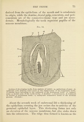
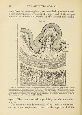
![dental ridge. From the inner side of this ridge epithelial cords or dental bulbs arise which will form the temporary teeth. These bulbs continue to grow into tlask-shaped bodies, their interior being mature cells, while externally they are covered with germinal cells. Later on, these become the enamel organs. About this time the fibrous tissue of mesoblastic origin begins to surround the enamel organs. The base of the enamel organ becomes indented, due to this papillary growth, and the future tooth is being shaped. The fibrous tissue which lies under the enamel organ is condensed and the bloodvessels begin to form. This fibrous tissue gradually surrounds the whole cord and is called the dental sac, which with its contents comprises the dental follicle. From the sides of the epithelial cords, buds develop forming the enamel organs for the permanent teeth. The cells of the enamel organ are now differentiated into several layers, an outer or cuboidal layer, and an inner or columnar layer, with a middle or stellate layer called the enamel jelly. The mesodermic ])apilhe which arc caj)pcd by the enamel organ now push toward the surface, their blood-supply being well established, while the cells of the surrounding connective tissue have elongated to form the odontoblasts, which later produce the dentine. Formation of Enamel.—Gradually globules of a calcareous nature, known as the enamel-prisms, are secreted by the inner columnar layer of cells of the enamel organ. Between these prisms the cementing substance is secreted, the deposit of enamel occurring from within out. Formation of Dentine.—The odontoblasts take from the blood the material for making the dentine, which they secrete, the same being deposited in layers from without in. The cementum has its origin from the dental sac, being a layer of bone adherent to the dentine and covering the fang. The central portion of the dentinal papillfB is changed into pulp after a sufficient amount of dentine has been deposited. The permanent teeth develop from the secondary enamel organs which are left as remnants of the original dental](https://iiif.wellcomecollection.org/image/b2805801x_0070.jp2/full/800%2C/0/default.jpg)