Normal histology : a manual for students and practitioners / by John R. Wathen.
- Wathen, John Roach, 1872-
- Date:
- [1905]
Licence: In copyright
Credit: Normal histology : a manual for students and practitioners / by John R. Wathen. Source: Wellcome Collection.
74/232
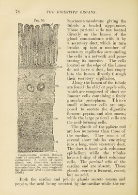
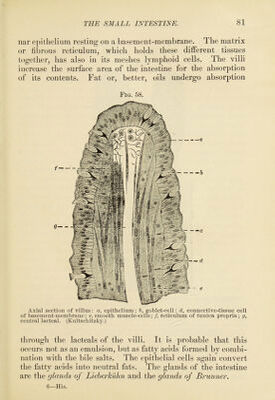
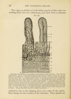
![Fio. 50. basement-membrane giving the tubule a beaded appearance. Those parietid cells not located directly on the lumen of the gland communicate with it by a secretory duct, which in turn breaks up into a number of secretory capillaries surrounding the cells in a network and pene- trating its interior. The cells located on the edge of the lumen do not have a duct, but empty into the lumen directly through their secretory capillaries. Along the lumen of the tubule are found the chief or peptic cellsj which are composed of short co- lumnar cells containing a finely granular protoplasm. These small columnar cells are sup- ])osed to secrete the digestive ferment pepsin, and also mucus, while the large parietal cells are the acid-forming cells. The glands of the pyloi'ic end are less numerous than those of the cardiac. They consist of several short tubules emptying into a long, wide excretory duct. The duct is lined with columnar epithelium while the tubules have a lining of short columnar cells. The parietal cells of the cardiac end are absent. These glands secrete a ferment, I'ennet, but no acid. Both the cardiac and ])yloric glands secrete mucus and pepsin, the acid being secreted by the cardiac while the en- Diagranimatic section through coats of stomach: m, mucosa; e, epithelium; d, outlet of peptic gland; mm, muscularis mucosfe; sm, submucosa; cm, inner circular muscular layer; Im, outer longitudi- nal muscular layer; s, peritoneum. (Mall.)](https://iiif.wellcomecollection.org/image/b2805801x_0074.jp2/full/800%2C/0/default.jpg)