Normal histology : a manual for students and practitioners / by John R. Wathen.
- Wathen, John Roach, 1872-
- Date:
- [1905]
Licence: In copyright
Credit: Normal histology : a manual for students and practitioners / by John R. Wathen. Source: Wellcome Collection.
79/232
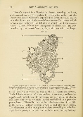
![found between the bases of the villi in the small intestine and also in the large intestine, in w somewhat longer. They are regnla ons row and have an ampnlla-like \ ^ The glands of Brunner are s})ecial secretory structures found in the dnodennm and first part of the jejunum. These are compound tubular or racemose glands lying in the snbrnncosa and discharging their secretion into a long, slender duct which opens between the bases of the villi. Sometimes these glands are also found in the pyloric end of the stomach. They are easily recognized because of their ])osition in the snbmncosa, and their ducts pass through the mnscnlaris mucosa. The cells of these glands are composed of finely granular cylindrical epithelium, and the extremities of the gland-tnbnles, being dilated, resemble alveolar glands. The submucous coat is made up of a loose areolar tissue, containing the larger blood- and lymj)h-vessels, the nerve- plexus of Meissner, and lymphatic glands. These lymphatic glands are of two kinds, the solitary glands and the agminated glands. The solitary glands are isolated lymph-follicles found lying in the submucosa throughout the entire intestine, but most numerous near the ileoc^ecal valve. They consist * of a dense reticular tissue, closely filled with lymph-cells. They have no ducts and communicate with the lacteal system through the aid of large lymph-spaces at their base, which are continuous with the interspaces of the retiform tissue. The agminated glands^ or Beyer’s patches, are large groups or aggregations of lymph-follicles, usually oval iu outline, from twenty to thirty in number, from a half to four inches in length, and situated in the submucosa of the intestine, especially in the lower part of the ileum. Their free surface is usually devoid of villi. The muscular coat consists of a thick inner or circular, and a thinner outer or longitudinal layer of involuntary muscle- fibre. Between these is the nerve-plexus of Auerbach. The fibroserous coat is similar to the same laver of the stomach, and is composed of fibroelastic tissue covered with the endothelial cells of the peritoneum. hich latter place they are rly arranged in a continu- vudeniniT of their lamina.](https://iiif.wellcomecollection.org/image/b2805801x_0079.jp2/full/800%2C/0/default.jpg)