A text-book of diseases of the nose and throat / by D. Braden Kyle.
- Kyle, D. Braden (David Braden), 1863-1916
- Date:
- 1899
Licence: Public Domain Mark
Credit: A text-book of diseases of the nose and throat / by D. Braden Kyle. Source: Wellcome Collection.
24/680 (page 22)
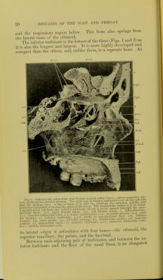
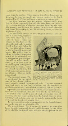
![nasal cavitv, extending some distance over each orbit, and giving rise to the prominences over the root of the nose. Uka the sphe- noidal sinuses, they develop with advancing age. Tliey communi- cate with the middle meatus by the infiindibulum, as already described. . , n n ^ A small sinus in the upper anterior part ot tlie antrum ot Hi-hmore has been observed. It is quite separate from the niax- ilkuy sinus, and through it runs the canal carrying the intra-or- bital nerve. It is well shown in Fig. 6. The maxillary sinuses (Fig. 6), or antra of Highmore, are two large pyramidal cavities situated one in the body of each superior maxillary bone. The roof of each antrum is formed by the floor of the orbit, its floor bv the alveolar process, its external wall by the facial surface, and its posterior wall by the zygomatic surface of the superior maxillary. It opens into the middle meatus (Fig. 2), near the posterior part of the hiatus semihmarif, by a circular opening, the ostiujn maxillare, behind which_ is occasionally a second' opening, the ostium maxillare accessoriii^. These cavities vary much in size, both in races and in individuals. They are frequently crossed by thin laminae of bone. In the pos- terior wall are the canals transmitting the posterior dental vessels and nerves to the teeth, and on the floor may often be found con- ical proiections caused by the roots of the first and second molar teeth In the anterior region of the inferior meatus is the orihce of the lacrimal or nasal duct, leading from the lacrimal sac to the nose (Fig. 114). . The mucous membrane lining the accessory sinuses dilierft slightly from the nasal mucous membrane. The epithelial lining consists in a single layer of pavement epithelial cells. The base- ment membrane and submucosa are much thinner than the exposed mucous surfaces, and the gland element largely limited to the ori- fice communicating with the nasal tract, the glands of the sinus mucous membrane being few in number. The bony walls of the nasal cavities and the accessory sinuses are completely lined by mucous membrane, which in front is con- tinuous with the skin, and at the posterior nares with the mucous membrane linimr the plmrynx. This membrane, which is vari- ously known as the pituitary or phlegm-producmg, the bclinei- derian, or the nasal mucosa, is intimately^applied to the bonv structure, varies in thickness and character in different areas, and modifies greatly the size of the nasal fossae and their accessory sinuses and orifices, as seen in the skull. It is thickest over the turbinated bones, somewhat thinner over the sep um am vei> thin over the floor, the under surfac(«s of the turbinated bones, and in the accessory cavities. . r . .if.,,. The (^olor of the nasal mucosa also varies. 1 n the up])er oi ollac - tory region, including the roof, superior turbinated bone, superior](https://iiif.wellcomecollection.org/image/b20388469_0024.jp2/full/800%2C/0/default.jpg)