Volume 1
Natural history of Victoria : prodromus of the zoology of Victoria; or figures and descriptions of the living species of all classes of the Victorian indigenous animals / by Frederick McCoy.
- Frederick McCoy
- Date:
- [1885-90]
Licence: Public Domain Mark
Credit: Natural history of Victoria : prodromus of the zoology of Victoria; or figures and descriptions of the living species of all classes of the Victorian indigenous animals / by Frederick McCoy. Source: Wellcome Collection.
Provider: This material has been provided by Royal College of Physicians, London. The original may be consulted at Royal College of Physicians, London.
506/630
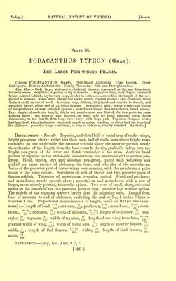
![This most beautiful Phasma is readily distinguished from the other two large species found near Melbourne, the Tropidoderus rhodomus and T. iodomus, figured in our Plates 69-70, by the beautiful rosy-pink colour of the membrane of the lower wings, with the bright rosy-carmine veins, and the same colour occupying as much of the basal portion of the costal area as can be covered over by the tegmina when at rest. It is also easily distinguished by the generic characters which separate the Podacanthi from the Tropidoderi, particularly the great length of the anal styles, the unkeeled tuberculated mesothorax, the three ocelli on the top of the head, and the undilated femora. M. Serville, in his “Histoire Naturelle des Insectes, Orthopteres,” p. 230, says that the antennae of the females are longer than the thorax ; but in our specimens they but slightly exceed the meta- thorax and mesothorax taken together, without the prothorax. If the basal joint be counted, there are 27 joints in the antennae, instead of 26, as given by Professor Westwood. The number of spines on the hind legs varies, but is usually about a dozen. I have not seen the males as yet, nor can I account for their apparent rarity. Specimens are in the collection from the Richmond Paddock and other localities near Melbourne. Explanation op Figures. Plate 80.—Fig. 1, female, natural size, in flying position. Fig. Ire, ditto, antenna;, head with the three ocelli, prothorax, and mesothorax, magnified. Fig. lb, ditto, side view of leg. Fig. 1c, ditto, side view of hind joints of abdomen, to show ovipositor. (N.B.—The young, with imperfectly developed wings, is figured in the resting position on Plate 79, fig. 3, and in it the ocelli are not visible.) Frederick McCoy. By Authority : John Ferres, Government Printer, Melbourne. [ 36 ]](https://iiif.wellcomecollection.org/image/b24757469_0001_0506.jp2/full/800%2C/0/default.jpg)