Affinity labelling and cloning of steroid and thyroid hormone receptors / edited by H. Gronemeyer.
- Date:
- [1988]
Licence: Attribution-NonCommercial-NoDerivatives 4.0 International (CC BY-NC-ND 4.0)
Credit: Affinity labelling and cloning of steroid and thyroid hormone receptors / edited by H. Gronemeyer. Source: Wellcome Collection.
24/332 page 20
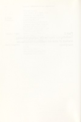
![20 Estrogen receptor eluting standard expressed as a percentage of the total activity in the chromatogram gives the radiochemical purity. Radiochemical purity analysis by TLC proceeds in a similar fashion, with the chromatogram being cut into 2-5 mm slices which are counted. Suitable chromatographic solvent systems are normal phase on silica gel (triethylamine-ethanol-ether: 10-20-70) and reversed phase on C-18 silica gel (water-acetonitrile: 30-70) (Simpson et al., 1987). П. COVALENT LABELING OF ESTROGEN RECEPTORS A. Labeling of cytosol receptors This protocol can be used in labeling cytosol receptors from a variety of tissue and cells. In tissue cytosols with high proteolytic activity, it may be necessary to include inhibitors such as molybdate (20 or 40 mM), leupeptin (Img/ml), phenylmethyl- sulfonylfluoride (PMSF, 1 mM), and soybean trypsin inhibitor (5 mg/ml) in order to obtain intact, undegraded receptor. The appropriate mixture and concentration of protease inhibitors will need to be determined for specific tissues. For studies with cytosol from immature (day 20-23) rat uterus or from MCF-7 cells, where proteolysis is low when preparations are maintained at 0-4°C, inclusion of 20 mM molybdate or 1 mg/ml leupeptin show >90% intact Mr 66000 receptor, but receptor degradation may be marked if preparations are incubated at higher temperatures (Monsma et al., 1984). Several buffer systems (based on Tris or phosphate) are appropriate for prep¬ aration of cytosol and labeling of receptors, and the choice of buffer may depend on the subsequent plans for the covalently labeled receptor. We have found TEG+Mo buffer, which contains 10 mM Tris-HCl, 1.5 mM EDTA, 10% (v/v) glycerol, and 40 mM sodium molybdate, pH 7.4 at 4°C, to be a good buffer for preparation of rat uterine or MCF-7 cell cytosol receptors. B. Typical protocol for labeling cytosol estrogen receptor The procedure given here describes the labehng of rat uterine estrogen receptors. It can be modified slightly by the investigator, as needed for other tissues. Uteri {ca 30 mg wet weight per uterus) are excised from 19-23 day old Sprague- Dawley Holtzman rats (Holtsman Co., Madison, WI) and are homogenized at 5 uteri/ml or TEG-I-Mo buffer {ca 5 mg soluble protein per ml). Homogenates are centrifuged at 180 OOOg for 30 min at 0-4°C, and the supernatant is carefully collected and then filtered through a Millipore 0.22 fxm filter to remove lipids. Dimethylforma- mide (DMF) is then added to give a final concentration of 3%. This uterine cytosol is then incubated for 2 h at 0°C with 20 nM [^H]Tam-Az or for 3 h at 15°C with 20 or 40 nM [^H]KN-Az in the absence and presence of a 100-fold excess of radioinert estradiol. (Cytosol labeling with [^H]KN-Az can also be carried out at 0°C for 3 h, but labeling is less efficient at the lower temperature; see Elliston et al., 1987). Ligands are prepared in ethanol solutions such that the final concentra¬ tion of ethanol does not exceed 1%. Labeling is terminated and excess Ugand removed by incubation with charcoal-dextran slurry (1 part slurry to 9 parts cytosol) for 10 min at 4°C, and charcoal is removed by centrifugation at 12000g (5 min), or samples are directly spotted onto filter paper disks and treated with organic solvents to measure covalent attachment (see below).](https://iiif.wellcomecollection.org/image/b18029310_0025.JP2/full/800%2C/0/default.jpg)


 No text description is available for this image
 No text description is available for this image
 No text description is available for this image