Affinity labelling and cloning of steroid and thyroid hormone receptors / edited by H. Gronemeyer.
- Date:
- [1988]
Licence: Attribution-NonCommercial-NoDerivatives 4.0 International (CC BY-NC-ND 4.0)
Credit: Affinity labelling and cloning of steroid and thyroid hormone receptors / edited by H. Gronemeyer. Source: Wellcome Collection.
266/332 (page 262)
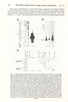
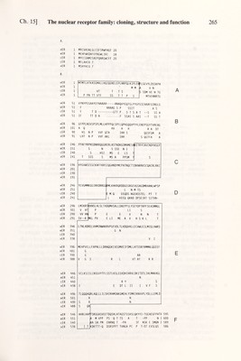
![The nuclear receptor family: cloning, structure and function [Ch. 15 The region containing the 5' end of the chicken progesterone receptor mRNA was first locahzed by Northern blotting of poly(A)^ RNA from laying-hen oviduct using the three single-stranded probes D, E and F described in Fig. 5C. Two clear bands of—4.5 and 3.3 kb and a fainter band of 6.5 kb were revealed using probes D SI NUCLEASE MAPPING NORTHERN ANALYSIS Ol X m I I probe A probe В probe С Primer probe D probe E probe F y/- УА 100bp I 1 Fig. 5 — Mapping of the chicken progesterone receptor mRNA 5' end. (A) A 6% sequencing gel showing si nuclease (lanes 3 and 4) and primer extension (lanes 1 and 2) mapping using poly(A)^ RNA from laying-hen oviduct (lanes 2 and 4) or from HeLa cells (lanes 1 and 3). The lanes CTAG show the sequence ladder obtained using the dideoxy method and oligo 2 as primer elongated in the presence of [a-^^S]dATP. Open triangles indicate the position of the major transcription start site. (B) Northern blot analysis using 15 /xg of RNA per slot. Lanes 1,3 and 5 consist of total HeLa cell RNA mixed with 2 ng of the Hindlll-PstI fragment (see (C)) as a positive control; lanes 2, 4 and 6 are poly(A)^ RNA from oviduct. The probes used for hybridization were D (lanes 1, 2), E (lanes 3, 4) and F (lanes 5, 6). (C) Map of the 5' region of the chicken progesterone receptor gene showing the probes and the Primer used in A and B. The M13 universal primer (open squares) was used to synthesize probes A and D, oligonucleo¬ tide 3 was used for the synthesis of probes В and E, oligonucleotide 2 for the synthesis of probe С and the Primer, and oligonucleotide 1 for probe F. The sequences of oligoneucleotides (1-3) indicated by the filled squares are depicted in Fig. 4. In all cases probe synthesis was carried out in the presence of [a-^ P]dATP, followed by digestion with Ncol (probes A, В and C), Bgll (probes D, E and the Primer) and Nsil (probe F) and purification. The Ncol site (marked with an asterisk) at -137 was introduced by site-directed mutagenesis. (From Gronemeyer et al. (1987).)](https://iiif.wellcomecollection.org/image/b18029310_0267.JP2/full/800%2C/0/default.jpg)