Affinity labelling and cloning of steroid and thyroid hormone receptors / edited by H. Gronemeyer.
- Date:
- [1988]
Licence: Attribution-NonCommercial-NoDerivatives 4.0 International (CC BY-NC-ND 4.0)
Credit: Affinity labelling and cloning of steroid and thyroid hormone receptors / edited by H. Gronemeyer. Source: Wellcome Collection.
273/332 (page 269)
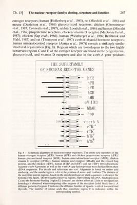
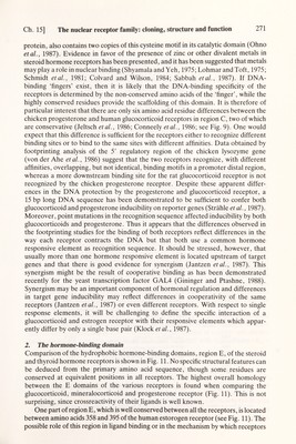
![Ch. 15] The nuclear receptor family: cloning, structure and function 269 genes present on the same chromosome. Furthermore, in a human hepatocellular carcinoma, the hepatitis В virus genome integrated into a gene, now called hap (de Thé et al., 1987), located on chromosome 3, which has sequence homology with region С of steroid hormone receptors and erb-A (Dejean et al., 1986). Thus, it is likely that there are additional members of this superfamily of transcriptional regulatory factors still to be isolated (see Fig. 10). 1. The DNA-binding domain Although the different steroid and thyroid hormone receptors trigger very different physiological and developmental programs, their putative DNA-binding domains (region C, see below) are highly homologous. At first sight, this may seem surprising, because it is this domain which interacts with specific promoter sequences of target genes, thus activating gene transcription (see below and Green and Chambón, 1987). Sequence analysis of the DNA-binding region of the steroid and thyroid hormone receptors shows, however, that at least part of this homology is apparently due to the requirement of a specific structural organization. The most striking feature of region С is the conserved arrangement of nine cysteines, which is common to all receptors as shown in Fig. 9. From this alignment, it is already apparent that this domain consists of two halves, each with a specific arrangement of cysteines. In fact, these halves are encoded in different exons in both the progesterone and the estrogen receptors (Jeltsch et al., 1986; unpublished work of our laboratory). The N-terminal half of domain С is characterized by the motif Cys-X2-Cys- Xi3-Cys-X2-Cys. This structure is reminiscent of the DNA-binding region of a class gal4 ppri argii tfiiia Consensus 11 21 VpN A l 1 l v vvte vv|c vvjc 'dIy GiD'E S'DE GDK DiK дк s!g sIg _ s|g!C hyg HYG HYG T[GlYjHYR tigIyIhyr tIgIyìhyr VWS VLT VLT С IT С I T С IT c,di ICIKR icjwt crlkklkcskekpk c'rlkkikcdqefps cirgrkvkcdlrhph aipf Vip F FF FF f F f f siqg-- amegq- avegq- t i QKNLHjPS tiqknlìhIpt tiqknlIhÌpt lknnwec aklevpc eksnlpc le;. , DGClDKRFTKK . , LK 'rIft. patnq agrnd agrnd kyegk kydgc tydgc T IiDK IVlDK I I|DK V IDK VI'DK V ijM NRjR|KS RiRiK N RlRiKN VTIR I TjR ITR QC ai LRlK LR¡K YRil fkik fk'k fk!k КС YEVGM. С Q AG M LQAGM lYVGM I SV'GM I SV'GM' adri repeat 98 CÍEV|CTRAFARQEHLK*RÍH'YR*S|H, Fig. 9 — Alignment of receptor DNA-binding domains. Shown are the human estrogen (hER), the chicken progesterone (cPR), and the human glucocorticoid (hGR) receptor together with human с-егЬ-.Л (hc-erbA), the chicken c-erb-A (cc-erb-A) and the v-erb-A protein. Boxed areas indicate complete amino acid identity between all the sequences, and dashes (—) indicate amino acid gaps for optimal alignment. Conserved cysteins are shaded. The Cys-X2-Cys-Xi3-Cys-X2-Cys motif present in the putative DNA-binding domain of the GAL4, PPRI and Argil yeast regulatory proteins is aligned with the corresponding motif of the steroid hormone receptors and erb A gene product. The consensus motif of the Xenopus transcription factor TFIIIA and the corresponding repeat 1 of the yeast regulatory proteins ADRI are also shown. The pairs of cysteine and histidine residues dictating the postulated 'finger' structures are boxed. Asterisks indicate positions where amino acid insertions may occur, and dots in the consensus sequence indicate variable residues. Numbers indicate the position of amino acid residues in each sequence.](https://iiif.wellcomecollection.org/image/b18029310_0274.JP2/full/800%2C/0/default.jpg)