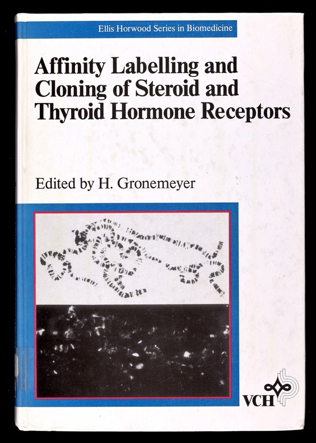
Affinity labelling and cloning of steroid and thyroid hormone receptors / edited by H. Gronemeyer.
- Date:
- [1988]
Licence: Attribution-NonCommercial-NoDerivatives 4.0 International (CC BY-NC-ND 4.0)
Credit: Affinity labelling and cloning of steroid and thyroid hormone receptors / edited by H. Gronemeyer. Source: Wellcome Collection.
38/332 page 34
![34 Glucocorticoid receptor [Ch. 2 homogenized in the presence of 2 mM Ca^^ (Fig. ЗА). However, receptors prepared by freeze-thaw lysis in buffer without Ca^^ (see Section III.A) exhibited no proteolytic fragments (Fig. 3B). D. Covalent labeling of rat liver receptors (Eisen et al., 1981) (NH4)2S04 precipitated liver cytosol (5 ml) was resuspended in 5 ml of TAPS buffer (25 mM TAPS, 50 mM NaCl, 20 mM Na2Mo04,10% (v/v) glycerol, pH=8.0 at 0°C) containing (1-2) X10^ M pH]Dex-Mes± 100-fold excess of [^H]dexamethasone for 2 h at 0°C. The yield of covalently labeled receptors is —80% of the total available specific pH]dexamethasone binding to cell-free receptors. Higher pH buffers can be used to increase the efficiency of covalent labeUng but the steroid binding activity of liver receptors decreases at higher pH so that the total yield of affinity labeled receptors also decreases (Simons et ai, 1983). E. Exchange binding 1. To detect preformed covalent complexes (Simons and Thompson, 1981) Crude HTC cell receptors were preincubated with 6.0x10^ M [^H]cortisol or 7.1 X10^ M [^H]Dex-Mes with or without 6 /х-М [^H]cortisol, for 3.2 h at 0°C (final protein concentration, 4.7 mg/ml). After addition of activated charcoal to remove free steroid, and to inactivate steroid-free receptors (Simons et al., 1980b), followed by centrifugation to pellet the activated charcoal, the preincubated cytosols were adjusted to 1.9xl0~® M pH]dexamethasone with or without 11 /мМ [^HJdexametha- sone. After subsequent incubation at 0°C, activated charcoal was again added to remove free steroid and the amount of specifically bound [^Hjdexamethasone (= total—non-specific binding) that was formed by exchange binding in cytosols prein¬ cubated with Cortisol, Dex-Mes, or Dex-Mes plus 85-fold excess Cortisol was determined. Maximal exchange binding of Dex-Mes pretreated receptors (~20°% of Cortisol pretreated receptors) was obtained after incubation for about 2 h. 2. Covalent labeling of previously bound receptors (Housley et al., 1985) L-cell cytosol (1.0 ml) containing 10 mM sodium molybdate and 2 mM dithiothreitol was stirred on ice for 2.5 h with 1/2 volume of deoxycorticosterone-agarose (Gran¬ dies et al., 1984) that was equiUbrated at 4°C in 10 mM Hepes, 10 mM molybdate and 2mM dithiothreitol, pH7.35. The agarose-bound components of cytosol were collected by centrifugation and washed six times with 10 volumes of cold Buffer A (10 mM Hepes, 10 mM molybdate, 10% (w/v) glycerol, 0.5 mM EDTA, pH7.6 at 4°C). Receptor was eluted from the resin by suspension in an equal volume (0.5 ml) of Buffer A containing 2.5 fjM pH]Dex-Mes and stirring on ice for 4-6 h. The suspension was centrifuged in a syringe containing a porous polyethylene disk to recover the eluate containing the affinity labeled receptors. IV. PHOTO AFFINITY LABELING OF CELL-FREE RECEPTORS While photoaffinity labeling of glucocorticoid receptors does not require prior protein purification, most of the reports have employed partially purified receptor (Westphal et al., 1981; Payvar et ai, 1983; Gronemeyer et al., 1983; Gehring and](https://iiif.wellcomecollection.org/image/b18029310_0039.JP2/full/800%2C/0/default.jpg)


 No text description is available for this image
 No text description is available for this image
 No text description is available for this image