Affinity labelling and cloning of steroid and thyroid hormone receptors / edited by H. Gronemeyer.
- Date:
- [1988]
Licence: Attribution-NonCommercial-NoDerivatives 4.0 International (CC BY-NC-ND 4.0)
Credit: Affinity labelling and cloning of steroid and thyroid hormone receptors / edited by H. Gronemeyer. Source: Wellcome Collection.
49/332 page 45
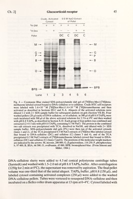
![Ch. 2] Glucocorticoid receptor 45 Crude, Activated 0.5 M NaCI Extract Lane 1 2 3 4 5 6 Fig. 4 — Coomassie Blue stained SDS-polyacrylamide slab gel of [^H]Dex-Mes±[^H]dexa- methasone labeled cytosol bound to DNA-cellulose or to cellulose. Crude UTC cell receptors were labeled with 1.6x10^M pH]Dex-Mes±1.6xlO~^M [^H]dexamethasone and then activated as described in Sections III.С and X.A. Aliquots of the activated solutions were diluted 1:2 with 2x SDS sample buffer for subsequent analysis on gels (Section VII.B). Four washed pellets (20 /xl each) of DNA-cellulose, or of cellulose, in 300 /х1 of pH 8.8 TAPSq were each incubated with 500 /х1 of the above activated solutions for 2.5 h at 0°C and then washed with pH 8.8 TAPSo as described in Section X.D. Each group of four pellets was combined and extracted (2x0.5 ml) with pH 8.8 TAPSq containing 0.5 M NaCl. The protein in the combined two salt extracts was precipitated with TCA, dissolved in NaOH, and diluted with 2x SDS sample buffer. SDS-polyacrylamide slab gels (9%) were then run of the activated cytosols (lanes 1 and 2), of the TCA precipitated 0.5 M NaCl extracts of [^H]Dex-Mes labeled cytosol that bound to DNA-cellulose (DC) and cellulose (C) (lanes 3 and 4), and of the TCA precipitated 0.5 M NaCl extracts of pH]dexamethasone labeled cytosol that bound to DNA- cellulose (DC) and cellulose (C) (lanes 5 and 6). The positions of the molecular weight markers are indicated by the arrows: M, myosin, 200 ООО; G, ß-galactosidase, 116 250; P, Phosphorylase b, 97 400; В, BSA, 66 300; О, ovalbumin, 45 ООО; ВРВ, bromophenol blue. (From Simons and Miller, 1984.) DNA-cellulose slurry were added to 4.5 ml conical polystyrene centrifuge tubes (Sarstedt) and washed with 1.5-2.0 ml of pH 8.8 TAPSq buffer. After centrifugation (1250g for 2 min at 0°C), the supernatant was removed by aspiration. The final pellet volume was one-third that of the initial aliquot. TAPSq buffer, pH 8.8 (150 /ttl), and labeled cytosol containing activated complexes (250 /xl) were added to the washed DNA-cellulose pellets. Tubes were vortexed to resuspend DNA-cellulose and then incubated on a Bélico roller drum apparatus at 13 фт at 0-4°C. Cytosol labeled with](https://iiif.wellcomecollection.org/image/b18029310_0050.JP2/full/800%2C/0/default.jpg)


 No text description is available for this image
 No text description is available for this image
 No text description is available for this image