Affinity labelling and cloning of steroid and thyroid hormone receptors / edited by H. Gronemeyer.
- Date:
- [1988]
Licence: Attribution-NonCommercial-NoDerivatives 4.0 International (CC BY-NC-ND 4.0)
Credit: Affinity labelling and cloning of steroid and thyroid hormone receptors / edited by H. Gronemeyer. Source: Wellcome Collection.
51/332 page 47
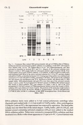
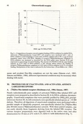
![Ch. 2] Glucocorticoid receptor 47 pHJdexamethasone was incubated with DNA-cellulose for 0.5-1.25 h; cytosol labeled with pH]Dex-Mes was incubated for 1-1.75 h. In both cases, >80% of the maximal binding is seen after 15 min and is stable for 1-4 h. Cytosol labeled with either steroid in the presence of excess [^H]Dex-Mes was incubated for 1.5-3 h. Binding was terminated at equilibrium by centrifugation (1250g for 2 min at 0°C) and the supernatant was removed by aspiration. A single wash of the DNA-cellulose pellets dramatically reduced the non-specific binding but had little effect on the observed specific binding; a second wash caused Httle further change and was omitted. Thus, the pellets were routinely washed with 1.0 ml of TAPSq buffer, pH 8.8, centrifuged as described above, aspirated to complete dryness with a flame- tapered Pasteur pipet, and incubated for at least 1 h at room temperature with 0.2 ml of pH 8.8 TAPSo buffer (to dissociate bound steroid and/or receptors) before being transferred to a scintillation vial with two 0.1 ml H2O washes and counted in 10 ml of Hydrofluor. Duplicates generally varied by less than 5%. Specific binding of [^H]steroid-receptor complexes to DNA-cellulose was calculated as the difference between binding of cytosol labeled in the absense (total binding) and presence (non¬ specific binding) of excess [^HJdexamethasone. Non-specific binding was usually less than 7% or 25% of the total binding in pH]dexamethasone, or pH]Dex-Mes labeled cytosols respectively. E. DNA competition binding assay (Miller etal., 1984) This assay is a modification of the above DNA pellet binding assay and can be used with non-covalent pHJdexamethasone, and covalent pH]Dex-Mes, receptor-ster¬ oid complexes. Activated pH]labeled receptor-steroid complexes (250)al) were incubated with 1 unit of DNA-cellulose (in 100 fxl of pH 8.8 TAPSq buffer; 1 unit of DNA-cellulose=33.3 ¡xX of DNA-cellulose pellet at a concentration of 1.2 mg DNA/ ml of cellulose) and 50 of TE buffer (10 mM Tris, 1 mM EDTA, pH 7.4 at room temperature) for control binding or with 0.625 units of DNA-cellulose (in 100 /xl of pH 8.8 TAPSo buffer) and soluble DNA (in 50 /xl of TE buffer) for 0.75-2.55 h at 4°C. Samples were processed as described above. Non-specific binding for each condition was determined by parallel incubations of the appropriate amounts of DNA-cellulose and soluble DNA with cytosol labeled in the presence of excess [^H]dexamethasone. Specific binding was calculated as the difference in the amounts of binding to DNA-cellulose by cytosols labeled in the absence and presence (i.e. non-specific binding) of excess [^H]dexamethasone. Specific binding in the presence of soluble DNA was then expressed as a percentage of specific binding to control (i.e. 1 unit of DNA-cellulose), and plotted as in Fig. 6. The theoretical basis of this assay is outlined elsewhere (Simons, 1977). F. DNA filter binding and footprinting These assays have not yet been described using affinity labeled glucocorticoid receptors. However, owing to the nearly idential cell-free DNA binding properties of non-covalent dexamethasone, and covalent Dex-Mes, receptor-steroid com¬ plexes (Simons et al., 1983; Simons and Miller, 1984, 1986; Miller etal., 1984), it is expected that very similar, if not identical, procedures can be used for non-covalent and affinity labeled complexes. Since the nuclear binding of non-covalent dexameth-](https://iiif.wellcomecollection.org/image/b18029310_0052.JP2/full/800%2C/0/default.jpg)


 No text description is available for this image
 No text description is available for this image
 No text description is available for this image