Affinity labelling and cloning of steroid and thyroid hormone receptors / edited by H. Gronemeyer.
- Date:
- [1988]
Licence: Attribution-NonCommercial-NoDerivatives 4.0 International (CC BY-NC-ND 4.0)
Credit: Affinity labelling and cloning of steroid and thyroid hormone receptors / edited by H. Gronemeyer. Source: Wellcome Collection.
52/332 page 48
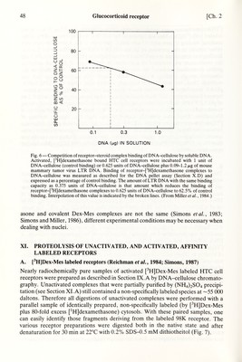
![48 Glucocorticoid receptor [Ch. 2 DNA (мд) IN SOLUTION Fig. 6 — Competition of receptor-steroid complex binding of DNA-cellulose by soluble DNA. Activated, [^H]dexamethasone bound HTC cell receptors were incubated with 1 unit of DNA-cellulose (control binding) or 0.625 units of DNA-cellulose plus 0.09-1.2 /xg of mouse mammary tumor virus LTR DNA. Binding of receptor-[^H]dexamethasone complexes to DNA-cellulose was measured as described for the DNA pellet assay (Section X.D) and expressed as a percentage of control binding. The amount of LTR DNA with the same binding capacity as 0.375 units of DNA-cellulose is that amount which reduces the binding of receptor-[^H]dexamethasone complexes to 0.625 units of DNA-cellulose to 62.5% of control binding. Inteфolation of this value is indicated by the broken hnes. (From Miller et al., 1984.) asoné and covalent Dex-Mes complexes are not the same (Simons et ai, 1983; Simons and Miller, 1986), different experimental conditions may be necessary when dealing with nuclei. XL PROTEOLYSIS OF UNACTIVATED, AND ACTIVATED, AFFINITY LABELED RECEPTORS A. [^H]Dex-Mes labeled receptors (Reichman etaL, 1984; Simons, 1987) Nearly radiochemically pure samples of activated [^H]Dex-Mes labeled HTC cell receptors were prepared as described in Section IX. A by DNA-cellulose chromato¬ graphy. Unactivated complexes that were partially purified by (NH4)2S04 precipi¬ tation (see Section XI. A) still contained a non-specifically labeled species at —55 ООО daltons. Therefore all digestions of unactivated complexes were performed with a parallel sample of identically prepared, non-speciñcally labeled (by [^H]Dex-Mes plus 80-fold excess [^HJdexamethasone) cytosols. With these paired samples, one can easily identify those fragments deriving from the labeled 98K receptor. The various receptor preparations were digested both in the native state and after denaturation for 30 min at 22°C with 0.2% SDS-0.5 mM dithiotheitol (Fig. 7).](https://iiif.wellcomecollection.org/image/b18029310_0053.JP2/full/800%2C/0/default.jpg)


 No text description is available for this image
 No text description is available for this image
 No text description is available for this image