Affinity labelling and cloning of steroid and thyroid hormone receptors / edited by H. Gronemeyer.
- Date:
- [1988]
Licence: Attribution-NonCommercial-NoDerivatives 4.0 International (CC BY-NC-ND 4.0)
Credit: Affinity labelling and cloning of steroid and thyroid hormone receptors / edited by H. Gronemeyer. Source: Wellcome Collection.
53/332 page 49
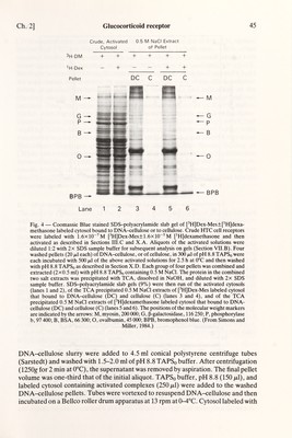
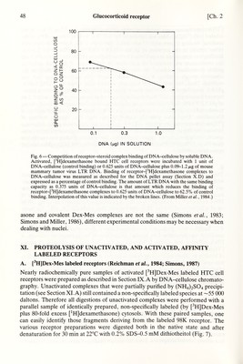
![Ch. 2] Glucocorticoid receptor 49 Native and denatured complexes were treated at 10°C for 20 min with enough stock solution (2.3 mg/ml in water) of trypsin (TPCK treated), chymotrysin (TLCK treated), or Staphylococcus aureus V8 protease to give final protease concentrations of 0.06-100 /Mg/ml. Digestions were stopped by freezing the samples in dry ice. For gel analysis, the samples were thawed just long enough to add 2x SDS sample buffer (0.6 M Tris (pH 8.85), 2% SDS, 0.2 M dithiothreitol, 20% (v/v) glycerol, and bromphenol blue) and refrozen at —78° until heated at 100°C for 5 min and then electrophoresed on 10.8% SDS-polyacrylamide gels. Limit protease digestion was achieved by treating denatured, [^H]Dex-Mes labeled complexes with 150 /xg/ml of protease for up to 48 h at room temperature. These digests were analyzed on 15% SDS-polyacrylamide gels, which resolve bands down to a molecular weight of ~1350 daltons (Simons, 1987). B. Proteolysis of photoafRnity labeled receptors (Gehring and Hotz, 1983; Gronemeyer eia/., 1983) Several proteases (trypsin, chymotrypsin, endoproteinase Lys-C, endoproteinase Arg-C) have been used to digest photoaffinity labeled receptors. Several different digestion conditions have been used. The main difference with digestion of Dex-Mes labeled receptors is that the concentration of photoaffinity labeled receptors is usually much lower. XII. LOCALIZATION OF MODIFIED AMINO ACID(S) IN AFFINITY LABELED RECEPTORS A. [^H]Dex-Mes labeled receptors (Simons eia/., 1987) Trypsin, chymotrypsin, and V8 protease limit digestion of nearly radiochemically pure samples of activated, [^H]Dex-Mes labeled HTC cell receptors in the denatured state was conducted as described in Section XL A. Efforts to purify a limit digest fragment for direct amino acid sequencing were thwarted by the extreme hydropho- bicity of these labeled fragments. Thus the [^H]-labeled, V8 limit digest fragment has an apparent Mr=1790±200 (SD, n = \l) on 15% SDS-polyacrylamide gels (Simons, 1987) and can be separated from most of the other proteins and peptides in the crude digestion mixture on 15% SDS-polyacrylamide gels but not on C3 HPLC columns. The limit digest fragment could not be eluted from Cis HPLC columns. Lyophiliza- tion of the digested sample caused most of the [^H] Dex-Mes labeled peptide to stick tightly to the walls of the tube and to resist resolubilization by most solvents. The labeled material was retained equally well by non-specific, and anti-dexamethasone, antibodies and stuck to Amicon 'Centricon 10' ultrafiltration membranes. All the radioactivity in crude V8 protease limit digests appeared in the void volume of Sephadex columns, regardless of whether or not the hmit digest solution was first heated at 100°C for 5 min before being chromatographed (a) on LH-20 columns with pH 8.8 TAPSo buffer containing 10 mM ß-mercaptoethanol, (b) on LH-20 columns with TAPSo-ß-mercaptoethanol buffer containing 4 M guanadinium hydrochloride, or (c) on either LH-20 or G-25 columns with TAPSq buffer containing 1% SDS and 0.1 M dithiothreitol. Good recoveries (60-70%) of the labeled material were obtained from Sep-pak](https://iiif.wellcomecollection.org/image/b18029310_0054.JP2/full/800%2C/0/default.jpg)


 No text description is available for this image
 No text description is available for this image
 No text description is available for this image