The surgical anatomy of inguinal herniæ, the testis and its coverings / by Thomas Morton.
- Morton, Thomas, 1813-1849.
- Date:
- 1841
Licence: Public Domain Mark
Credit: The surgical anatomy of inguinal herniæ, the testis and its coverings / by Thomas Morton. Source: Wellcome Collection.
67/134 (page 269)
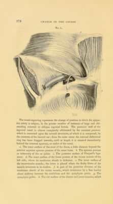
![CToacliccl upon by tlic hernial tumour, and its lower edge becomes in consequence rather more arched than ordinary, and the conjoined tendons of the two muscles (the internal oblique and trailsvcrsalis),—where they descend together to be inserted into the inner extremity of the ilco-pcctincal line of the os pubis, and form the inner part of the posterior wall of the inguinal canal,—are displaced more or less inwards, nearer to the outer edge of the rectus muscle ; so that instead of extend- ing an inch, and even more than this, from the edge of that muscle, they do not reach more than a (luartcr of an inch, and frequently even a less distance from it than this. In the healthy condition of these parts, the conjoined tendons consti- tute a thin and flat band of fibres; but, when an oblique inguinal hernia has existed for a considerable period, and, at the same time, forms a large protrusion, they arc no longer able to resist the tendency which the weight of the contents of the hernial sac has to drag inwards that portion of the posterior wall of the inguinal canal, which is composed of the fascia transvcrsalis and the conjoined tendons, and hence they give way, and are pushed aside in the direction of the mesial line of the boily, and so change their extended form for that of a thick, narrow, and strong band. (See Plate V. figs. 1 and 2, and woodcut No. 5, figs, f, m.) Such of the fibres of the internal oblique muscle as take their oiigin fiom the middle portion of Poupart’s ligament arc more or less displaced by the hernial sac as it descends through the inguinal canal; so that they no longer run, in an oblique^direc- tion, downAvards and inwards to their attachment to the os pubis, but arc forced upwards by the rounded anterior surface of the sac, as it protrudes between the lower edge of the muscle and the spermatic con], and are so made to form a thick mus- cular arch, the concavity of Avhich embraces about two-thirds of the circumference of the tumour as it lies within the iimuinal canal. ® Tlic cremaster muscle is frequently found very mucli altered rom Its natural and healthy condition, particularly if the hernia should be large and of long duration. The fibres of this muscle may be enlarged to throe or four times their ordinary thickness losing, at the same time, much of their muscular character, and t I'x/e Plale HI. fig, r, and woodcut No. 3, at page 2d(i, figs. i,g.](https://iiif.wellcomecollection.org/image/b22005092_0067.jp2/full/800%2C/0/default.jpg)