Diseases of the nose and its accessory cavities.
- Watson, W. Spencer (William Spencer), 1836-1906.
- Date:
- 1875
Licence: Public Domain Mark
Credit: Diseases of the nose and its accessory cavities. Source: Wellcome Collection.
Provider: This material has been provided by the Augustus C. Long Health Sciences Library at Columbia University and Columbia University Libraries/Information Services, through the Medical Heritage Library. The original may be consulted at the the Augustus C. Long Health Sciences Library at Columbia University and Columbia University.
96/506
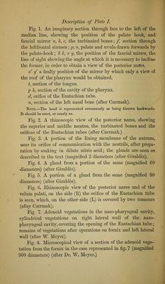
![DesGTvgtion of Plate I. Fig. 1. An imaginary section throngli face to the left of the median line, shomng the position of the palate hook, and faucial mirror a, h, c, the turbinated bones; f, section through the leftfrontal sinuses ; jp, u, palate and uvula drawn forwards by the palate-hook; hk, x y, the position of the faucial mirror, the line of sight showing the angle at which it is necessary to incline the former, in order to obtain a view of the posterior nares. X y a faulty position of the mirror by which only a view of the roof of the pharynx would be obtained. t, section of the tongue, ]) A, section of the cavity of the pharynx. d, orifice of the Eustachian tube. n, section of the left nasal bone (after Czermak). !NOTE.—The liead is represented erroneously as being tlirown backwards. It sbould be erect, or nearly so. Fig. 2. A rhinoscopic view of the posterior nares, showing the superior and middle meatus, the turbinated bones and the orifices of the Eustachian tubes (after Czermak.) Fig. 3. A portion of the lining membrane of the antrum, near its orifice of commimication with the nostrils, after prepa- ration by soaking in dilute nitric acid; the glands are seen as described in the text (magnified 2 diameters (after Giraldes). Eig. 4, A gland from a portion of the same (magnified 60 diameters) (after Giraldes). Eig. 5. A portion of a gland from the same (magnified 80 diameters) (after Giraldes). Eig. 6. Ehinoscopic view of the posterior nares and of the velum palati, on the side (E) the orifice of the Eustachian tube is seen, which, on the other side (L) is covered by two tumours (after Czermak). Eig. 7. Adenoid vegetations in the naso-pharyngeal cavity, cylindrical vegetations on right lateral wall of the naso- pharyngeal cavity, covering the opening of the Eustachian tube; remains of vegetations after operations on fornix and left lateral wall (after W. Meyer). Eig. 8. Microscopical view of a section of the adenoid vege- tation from the fornix in the case represented in fig. 7 (magnified 300 diameters) (after Dr. W. Meyer.)](https://iiif.wellcomecollection.org/image/b21204561_0096.jp2/full/800%2C/0/default.jpg)