Volume 1
The physiological anatomy and physiology of man / by Robert Bentley Todd and William Bowman.
- Date:
- 1845-1856
Licence: Public Domain Mark
Credit: The physiological anatomy and physiology of man / by Robert Bentley Todd and William Bowman. Source: Wellcome Collection.
Provider: This material has been provided by the Royal College of Physicians of Edinburgh. The original may be consulted at the Royal College of Physicians of Edinburgh.
419/476
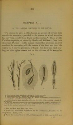
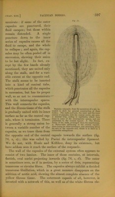
![11.4 lip municatc: it’ «oine of the outer capsules are punctured, their fluid escapes; but those within remain distended. A single puncture dowu to the inner series of capsules causes all the fluid to escape, and the whole to collapse ; and again, the cap- sules may he often peeled ofl in succession, shewing their union to be but slight. In fact, ex- cept by the few bands already mentioned, they are united only along the stalk, and for a vari- able extent at the opposite end. 'Pile stalk seems to be inserted into a kind of conical tube, which |>enetrates all the capsules in succession, but has its proper wall, so as not to communicate — with the intercapsular spaces. This wall connects the capsules, and the fibrous tlSSUO ot the stalk P»f mum roqwu'l'. from the U€*enterv at a cat: m. i» gradually unit'd with its inner & SZ surtdec m far M the central cap- = a,,]., wliorf. il ♦ There 1<M,P* ,n **>mr ,,f Ar '‘rrmtwidnr »rwwr», and ono suit, wnen ll lerinmaies. llltrt to the central e*),sul'. 4. The fii.rouu nonnrullv „ wtrnmr union of tl>' »taik. prolonged from the neurilemma. is generally a strong union ne- „ Nem..tut* .dAncing to the central c*P.aie, tween rt vnrinhle innnlter of the ‘htre st» *•«««i'»t»nce, ami .trctchintr alon? mien a \uriauie uumoer oi uit th, „„ to thf. oppositeend, .here it i» fired by » capsules, as we trace them from ,u,'erruUrenlargement, the opposite end of the central capsule towards the surface (fig. 76, b, e) ; this was called by Pacini the intercapsular ligament. We do not, with llenle and Kblliker, deny its existence, but have seldom seen it reach the surface of the corpuscle. The wall of the capsules of the external system often appears to consist of two laminar. The inner of these contains, at intervals, flattish, oval nuclei projecting inwards (fig. 76, a, d). The outer is sometimes seen, as if in section, by a series of dots, representing transverse or circular fibres. The capsules always exhibit a decided transverse fibrillation, which in a great measure disappears on the addition of acetic acid, shewing the almost complete absence of the yellow fibrous tissue. The outermost capsule of all, however, is invested with a network of this, as well as of the white fibrous ele-](https://iiif.wellcomecollection.org/image/b28043327_0001_0419.jp2/full/800%2C/0/default.jpg)