Stöhr's histology : arranged upon an embroyological basis / by Dr. Frederic T. Lewis ... From the 12th German ed. by Philipp Stöhr ... 6th American ed., with 450 illustrations.
- Philipp Stöhr
- Date:
- 1906
Licence: Public Domain Mark
Credit: Stöhr's histology : arranged upon an embroyological basis / by Dr. Frederic T. Lewis ... From the 12th German ed. by Philipp Stöhr ... 6th American ed., with 450 illustrations. Source: Wellcome Collection.
Provider: This material has been provided by the Augustus C. Long Health Sciences Library at Columbia University and Columbia University Libraries/Information Services, through the Medical Heritage Library. The original may be consulted at the the Augustus C. Long Health Sciences Library at Columbia University and Columbia University.
30/452 (page 18)
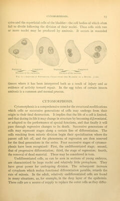
![II. GENERAL HISTOLOGY. HISTOGENESIS. Segmentation and the Formation of the Germ Layers. The body is composed of groups of similarly differentiated cells, similar therefore in form and function. Such groups are knov^Ti as tissues. Histology (Greek, ''(^roc, a textile fabric) is the science of tissues, and histogenesis deals with their origin. There are as many tissues in the body as there are sorts of substance; thus the liver consists essentially of hepatic tissue, and the bones of osseous tissue. All of these, however, are modifications of a small number of fundamental tissues, the histogenesis of which may now be considered. It has already been noted that a new human individual begins existence as a single cell, the fertihzed o\aim, formed by the fusion of two mature sexual cells, the spermatozoon and o\aim respectively. The fertilized ovum then divides by mitosis into a pair of cells. Fig. 19, A; these again divide making a group of four. Fig. 19, B; by repeated mitosis a mulberry- like mass of cells results, called the morula, Fig. 19, C. Development to this point is known as the segmentation of the ovum. A section through the morula is shown in D. An outer layer of cells surrounds the inner cell mass. Soon a cup-shaped cleft, crescentic in vertical section, forms between the outer and inner cells as shown in E, and this enlarges until the entire structure becomes a single-layered, thin-walled vesicle, within and attached to one pole of which is the inner cell mass. This mass gradually spreads beneath the outer layer until it forms a complete lining for the vesicle, which becomes consequently two layered, Fig. 19, G. The inner layer is called entoderm, and the outer layer, ectoderm.* The entire embryonic structure at this stage is called a blastodermic vesicle. On the upper surface of the vesicle the future -axis of the embryo is indicated by a thickened streak called the primitive streak. In front of this there is a groove in the ectoderm, also in the axial Hne of the future body. It is named the medullary groove, and just beneath it is a rod * The ectoderm is in part derived from the superficial cells of the inner cell mass, and in part from the primary outer layer of the vesicle. The former portion is to cover the body of ^the embryo, and the latter [named trophoblast] covers the fetal membranes. These membranes are to be described in a later chapter. They are omitted in the diagram? of Fig. xg.](https://iiif.wellcomecollection.org/image/b21206375_0030.jp2/full/800%2C/0/default.jpg)