Relations of diseases of the eye to general diseases : forming a supplementary volume to every manual and text-book of practical medicine and ophthalmology / Ed. by Henry D. Noyes.
- Knies, Max.
- Date:
- 1895
Licence: Public Domain Mark
Credit: Relations of diseases of the eye to general diseases : forming a supplementary volume to every manual and text-book of practical medicine and ophthalmology / Ed. by Henry D. Noyes. Source: Wellcome Collection.
Provider: This material has been provided by the Harvey Cushing/John Hay Whitney Medical Library at Yale University, through the Medical Heritage Library. The original may be consulted at the Harvey Cushing/John Hay Whitney Medical Library at Yale University.
33/492 page 15
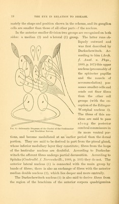
![adults. In the new-born the conditions are entirely different. In them it is evident that many combinations of fibres, which exist in the adult, have not yet developed. The investigations of Bern- heimer showed that in the new-born the optic nerve and chiasm contain only a limited number of medullated fibres. According to Flechsig's well-known assumption, they are only partly capable of function. If one eye or both eyes are destroyed at this period the degenerations are much more extensive. The ganglion cells of the primary optic ganglia degenerate almost completely. In the cortex of the occipital lobe much less is noticeable, very probably because, at this time, it is insufficiently connected with the primary optic ganglia and to a certain extent is still indifferent. In the further development of the brain the occipital cortex appears to assume other functions, probably on account of the more marked development of the system of association fibres, and the entire distribution of the cortical regions becomes different. It is probable that the auditory and particularly the tactile sense acquires a much larger cortical area. But these are merely assumptions which follow in part from the observations of cases of congenital blindness. To make a brief resume, we find in the optic nerve two kinds of fibres in approximately equal numbers: a, narrow centripetal axis cylinders from the cells of the ganglionic layer of the retina, which terminate, in great part, in a fine network in the three primary optic ganglia; and b, thicker centrifugal axis-cylinder processes from the ganglion cells of the anterior corpora quadrigemina, which spread out in the internal granular layer of'the retina. The remaining ganglion cells of the primary optic ganglia send their axis-cylinder processes (in the posterior third of the posterior limb of the internal capsule, immediately adjacent to the centripetal sensory tracts of the corona radiata) to the cortex of the occipital lobe, especially to the cuneus. The association fibres of the occipital cortex mainly pass forward. [The investigations of Henschen (Klinische und ana- tomische Beitrage zur Pathologie des Gehirns, Upsala, 1892, zweiter Theil) have narrowed the region of primary and direct corti- cal visual impressions to the calcarine fissure which is the inferior boundary of the cuneus.—N.]](https://iiif.wellcomecollection.org/image/b21017505_0033.jp2/full/800%2C/0/default.jpg)