Kirkes' handbook of physiology. / By W. Morrant Baker and Vincent Dormer Harris.
- William Senhouse Kirkes
- Date:
- 1888
Licence: Public Domain Mark
Credit: Kirkes' handbook of physiology. / By W. Morrant Baker and Vincent Dormer Harris. Source: Wellcome Collection.
Provider: This material has been provided by Royal College of Physicians, London. The original may be consulted at Royal College of Physicians, London.
110/912 page 86
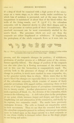
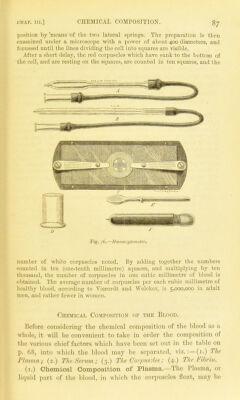
![Action of re-agents upon the colourless corpuscles.— Feeding the corjmscles.—If some fine pigment grauules, e.g., powdered vermilion, be added to a fluid containing colourless blood-cor- puscles, on a glass slide, these will be observed, under the micro- scope, to take up the pigment. In some cases colourless corpuscles have been seen with fragments of coloured ones thus embedded in their substance. This property of the colourless corpuscles is especially interesting as helping still further to connect them with the lowest forms of animal life, and to connect both with the organized cells of Avhich the higher animals are composed. The property which the colourless corpuscles ]30ssess of passing through the walls of the blood-vessels will be described later on. Enumeration of the blood-corpuscles.—Several methods are employed for counting the blood-corpuscles, most of them depending upon the same principle, i.e., the dilution of a minute volume of blood with a given volume of a colourless solution similar in specific gravity to blood plasma, so that the size and shape of the corpuscles is altered as little as possible. A minute quantity of the well-mixed solution is then taken, examined under the microscope, either in a flattened capillary tube (Malassez) or in a cell (Hayem & Nachet, Gowers) of known capacity, and the number of cor- puscles in a measured length of the tube, or in a given area of the cell is counted. The length of the tube and the area of the cell are ascertained by means of a micrometer scale in the microscope ocular ; or in the case of Gowers' modification, by the division of the cell area into squares of known size. Having ascertained the number of corpuscles in the diluted blood, it is easy to find out the number in a given volume of normal blood. Gowers' modification of Hayem & Nachet's instrument, called by him Hcemacyto- meter, appears to be the most convenient form of instrument for counting the corpuscles, and as such will alone be described (fig. 76). It consists of a small pipette (a), which, when filled up to a mark on its stem, holds 995 cubic millimetres. It is furnished with an india-rubber tube and glass mouth-piece to facilitate filling and emptying; a capillar}' tube (b) marked to hold 5 cubic millimetres, and also furnished with an india-rubber tube and mouth-piece ; a small glass jar (D) in which the dilution of the blood is performed ; a glass stirrer (b) for mixing the blood thoroughly, (f) a needle, the length of which can be regulated by a screw ; a brass stage plate (C) carrying a glass slide, on which is a cell one-fifth of a millimetre deep, and the bottom of which is divided into one-tenth millimetre squares. On the top of the cell rests the cover-glass, which is kept in its place by the pres- sure of two springs proceeding from the stage plate. A standard saline solution of sodium sulphate, or similar salt, of specific gravity 1025, is made, and 995 cubic millimetres are measured l)y means of the pipette into the glass jai-, and with this five cubic millimetres of blood, obtained by pricking the finger with a needle, and measured in the capillary pipette (B), are thoroughly mixed by the glass stirring-rod. A drop of this diluted blood is then placed in the cell and covered with a cover-glass, which is fixed in](https://iiif.wellcomecollection.org/image/b24757226_0110.jp2/full/800%2C/0/default.jpg)
 No text description is available for this image
 No text description is available for this image
 No text description is available for this image