Kirkes' handbook of physiology. / By W. Morrant Baker and Vincent Dormer Harris.
- William Senhouse Kirkes
- Date:
- 1888
Licence: Public Domain Mark
Credit: Kirkes' handbook of physiology. / By W. Morrant Baker and Vincent Dormer Harris. Source: Wellcome Collection.
Provider: This material has been provided by Royal College of Physicians, London. The original may be consulted at Royal College of Physicians, London.
57/912 page 33
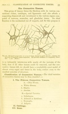
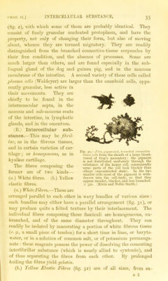
![CHAP. II.] CLASSIFICATION OF CONNECTIVE TISSUES. The Connective Tissues. This group of tissues forms the Skeleton with its various con- nections—bones, cartilages, and ligaments—and also affords a supporting framework and investment to the various organs com- posed of nervous, muscular, and glandular tissue. Its chief function is the mechanical one of support, and for this purpose it Fig. 28.—Horizontal preparation of cornea of fi'og, staiued in gold chloride ; showing the network of branched cornea corpuscles. The ground substance is completely colour- less. X 400. (Klein.) is so intimately interwoven with nearly all the textures of the body, that if all other tissues could be removed, and the con- nective tissues left, we should have a wonderfully exact model of almost every organ and tissue in the body, correct even to the smallest minutia; of structure. Classification of Connective Tissues.— The chief varieties of connective tissues may be thus classified :— I. The Fibrous Connective Tissues. A.—Chief Forms, a. White fibrous. h. pjlastic. c. Areolar. B.—Special Varieties. a. Gelatinous. b. Adenoid or Retiform. c. Neuroglia. , Adipose. II. Cartilage.](https://iiif.wellcomecollection.org/image/b24757226_0057.jp2/full/800%2C/0/default.jpg)
 No text description is available for this image
 No text description is available for this image
 No text description is available for this image