Icones obstetricae. A series of sixty plates illustrative of the art and science of midwifery, in all its branches / by A.L. Moreau ; edited with practical observations and tables, [by] J.S. Streeter.
- François-Joseph Moreau
- Date:
- 1842
Licence: Public Domain Mark
Credit: Icones obstetricae. A series of sixty plates illustrative of the art and science of midwifery, in all its branches / by A.L. Moreau ; edited with practical observations and tables, [by] J.S. Streeter. Source: Wellcome Collection.
19/206 page 1
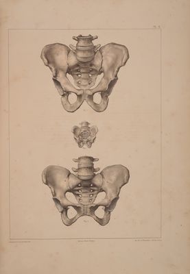
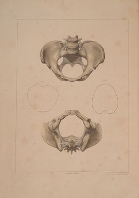
![OS SACRUM. Figure 1.—Anterior aspect. Figure 2.—Posterior aspect. Figure 3.—Lateral aspect. Figure 4.—Base. From a toa’. Fig. 1. The five bodies of the sacral vertebree forming so many superficial transverse sulci or depressions. From b' to b*. Fig. 1. The four transverse prominencies indicating the junctions of the bodies of the sacral vertebra. From c! to c’. Fig. 1. The four anterior sacral foramine. d. Fig. 1, 3, 4. The sacro-vertebral articular surface. e. Fig. 1, 2, 3, 4. The articular processes. f, Fig. 1, 3. Apex of the sacrum articulating with the base of the os coccygis. From g' to g*. Mig. 2. bral spinous processes. From h' to h‘. Fig. 2. The four posterior sacral foramine. i. Fig. 1, 4. The superior orifice of the sacral canal. j. Fig. 2. The inferior orifice of the sacral canal. k, k. Fig. 2. Cornua of the sacrum. They bound the termination of the The sacral spines in continuation of the verte- sacral canal laterally. ]. Fig. 3. Ear-shaped surface articulating with the os ilium. m,m. Fig. 2. Sacral groove, gutter or furrow. OS COCCYGIS. Fig. 5. Posterior surface. 1, 2, 3, 4. The four pieces of the os coccygis. a,a. Fig. 5. The coccygeal cornua. b, b. Tubercles of the coccyx. Final rudiments of the vertebral apo- physes. OS INNOMINATUM. Figure 6, 7. Yixternal or femoral surface. Figure 8. Internal or pelvic surface. a. Mig. 6,7. External iliac fossa. From b to c. Fig. 6, 7, 8. Crista ilii. From b to d. Fig. 6, 7, 8. Notch which separates the anterior and su- perior spinous process of the ilium from the anterior and inferior spinous process of the same bone. From c to e. Fig. 6, 7, 8. Notch which separates the posterior and supe- rior iliac spine from the posterior and inferior iliac spine. From e to f. Mig. 7, 8. Great sciatic notch. From f to g. Mig. 7, 8. Small sciatic notch. From g toh. Mg. 6, 7. Tuberosity of the ischium. From h to j. Fig. 6, 8. Ascending ramus of the ischium. From j to k. Fig. 6, 8. Descending ramus of the pubis. From k to p. Fig. 6, 8. Horizontal ramus or body of the pubis. a. Fig. 6, 7. External iliac fossa. b. Fig. 6, 7, 8. Anterior and superior spine of the ilium. c. Fig. 6, 7, 8. Posterior and superior spine of the ilium. e. Fig. 6, 7, 8. Posterior and inferior spine of the ilium. f. Fig. 7, 8. Sciatic spine. g,h. Fig. 6, 7, 8. Sciatic tuberosity. j. Fig. 6, 8. Point of union of the ascending ramus of the ischium and descending ramus of the pubis. k. Fig. 6, 8. Angle of the pubis. ]. Fig. 6, 8. Spine of the pubis. m. Fig. 6, 8. Pubic articular surface. n. fg. 6, 8. Thyroid or obturator foramen. o. Fig. 6, 7. Acetabulum. p. Fig. 6, 8. Tlio-pectineal eminence. q. Fig. 8. Brim of the pelvis, or prominent ridge which bounds the internal iliac fossa inferiorly and concurs in forming the abdomi- nal inlet (brim) of the pelvis. r. Mig. 8. Internal iliac fossa. s. fig. 8. Articular surface of the ilium, which, with the sacrum, forms the sacro-iliac symphysis or synchondrosis. t. Fig. 8. Obturator notch for the transmission of the obturator ves- sels and nerves. |](https://iiif.wellcomecollection.org/image/b33545960_0019.jp2/full/800%2C/0/default.jpg)


 No text description is available for this image
 No text description is available for this image
 No text description is available for this image