Our teeth, how built up, how destroyed, how preserved / described and illustrated by R. Denison Pedley, and Frank Harrison.
- Pedley, Richard Denison.
- Date:
- 1908
Licence: In copyright
Credit: Our teeth, how built up, how destroyed, how preserved / described and illustrated by R. Denison Pedley, and Frank Harrison. Source: Wellcome Collection.
Provider: This material has been provided by The University of Leeds Library. The original may be consulted at The University of Leeds Library.
20/108 page 16
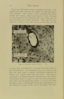
![Fig. 2 is an illustration of bone magnified 230 times. The gelatine has been removed by boiling and only the frame- work remains. The central cavity contained the blood vessels and nerves. Layer upon layer of celis s. round it, and extremely fine channels, like wavy lines, may be seen traversing the whole structure. This may be com- pared to a section across the root of a tooth (fig. 23), and F']g. 2.—Transverse Section ot Bone. it shows how nourishment is conveyed from the centre to the circumference. On referring to fig. 22 it will be seen that the tooth pulp is situated immediately beneath the dentine and enamel which form the crown. This pulp is composed of a delicate network of blood vessels and nerves. Surrounding them is a layer of cells, and fine prolongations from these run throughout the dentine or ivory of the tooth. Sensation—pain—is transmitted from these prolongations to the filaments of nerve in the root of the tooth; these fila-](https://iiif.wellcomecollection.org/image/b21506474_0020.jp2/full/800%2C/0/default.jpg)