Our teeth, how built up, how destroyed, how preserved / described and illustrated by R. Denison Pedley, and Frank Harrison.
- Pedley, Richard Denison.
- Date:
- 1908
Licence: In copyright
Credit: Our teeth, how built up, how destroyed, how preserved / described and illustrated by R. Denison Pedley, and Frank Harrison. Source: Wellcome Collection.
Provider: This material has been provided by The University of Leeds Library. The original may be consulted at The University of Leeds Library.
73/108 page 69
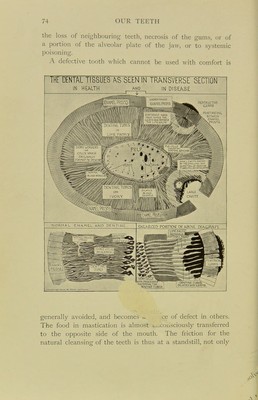
![Owing to the exposed condition of the teeth the physical •signs of caries or decay are easily observed, and reference to the following diagrammatic representations of the pro- gressive stages of tooth destruction will enable the reader DIAGRAM opthePHENOMENAopDENTALCARIES. SECTION OF TOOTH WITH 5LIRF?0UMDINC TISSUES IN SITU FiC 1. NORMAL TOOTH T155UES WITH COnrlENCINC CARIES AT A. Ftcll. CAVITVformeo THROUCH ' ENAMEL INTO DENTINE BY HEANS OF ACID BACTERIA.IrRITATINcPULP ANDCAUSINcSwELLlNG oftheBLOODI VESSELS iNFunnATioN andPAIN Ficlll. DEATH OF THE BLOODVESSEL AMD INFECTION OF THE PULPaVITI' WITH SEPTIC CERrlS FROnTKEnOUTH. VESSELS AROUND RAISING ' TOOTH IN SOCKET.Pain om biting Fic IV. OPENING INTO PULP cavity WITH FOOD OR DEBRIS ; PREVENTINC ESCAPE OF DECOHPOSINC '. CA5ES AT A AND FORCING A FASSJCE ■lUFLAMED] AtB FORMS AN ABSCESS WHICH 1^^ DiscwfiCEs AT Gas A CUn BOIL . Fig. 22 to recognize not only the results of such destruc-tion, but the necessity for treatment in the early stages. A typical tooth has been chosen, that of a premolar or bicuspid with a single root. The sections of it are vertical, and .show the whole tooth with the surrounding tissues //i situ. The first change is seen in the enamel (fig. 22, I, a), ■which loses its semi-translucent appearance at one spot, and](https://iiif.wellcomecollection.org/image/b21506474_0073.jp2/full/800%2C/0/default.jpg)