Plates of the arteries of the human body / after Frederic Tiedemann ... Engraved by E. Mitchell ... the explanatory references translated from the original Latin, with additional notes, by Dr Knox.
- Friedrich Tiedemann
- Date:
- 1835
Licence: Public Domain Mark
Credit: Plates of the arteries of the human body / after Frederic Tiedemann ... Engraved by E. Mitchell ... the explanatory references translated from the original Latin, with additional notes, by Dr Knox. Source: Wellcome Collection.
Provider: This material has been provided by The University of Glasgow Library. The original may be consulted at The University of Glasgow Library.
25/188 (page 3)
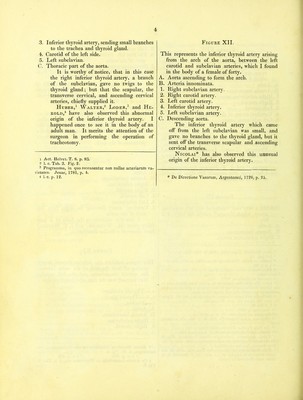
![FIGURE VIII. This shews an unusual origin of the left verte- bral artery from the arch of the aorta be- tween the left carotid and subclavian arteries, which I saw in the body of a man of fifty. A. Aorta. B. Arteria innominata. 1. Right subclavian artery. 2. Right carotid. 3. Left carotid. 4. Vertebral artery of the left side. 5. Left subclavian artery. C. Descending aorta. This variety very frequently occurs. It has been observed by J. Gr. de Bergen,1 Petsche,2 Trew,3 Loeseke,4 Boehmer,5 Morel,6 Huber,7 Morgagni,8 Winslow,9 Barbaut,10 Mekel,11 Sandifort,12 Nie- meyer,13 14 and J. F. Meckel the grandson.11 I have seen this anomaly five times in males as well as in females. Figure IX. Exhibits a very rare variety of the left vertebral artery, which Huber* saw in the body of a boy. A. Aorta. B. Arteria innominata. 1. Right subclavian artery. 1 Acta Eruditor. Lips. Ann. 1698, p. 295. Tab. 7. 2 1. c. pag. 14. •T Commerc. Liter. Norimberg. Ann. 1737, p. 185, No. 2. 4 1. c. p. 26. 5 Observation. Anat. Rarior. Fasc. 1. p. 11. No. 15. In a female body. 6 Journal de Medicine, 1757. Dec. 7 1. e. He saw this structure in a new born infant, and in one fifteen days old, in a boy of one year, in a girl of two, and another of five years old, and in an old woman. 8 De Sedib. et Causis Morbor. Epist. 3. Art. 20, 21. Epist. 15, Art. 26. Epist. 56. Art. 10. and 21. 9 1. c. No. 21. 10 Angiolog. p. 387. 11 Epistolae ad HalJerum. T. 2. p. 258. ]2 Observat. Anat. Patholog. Lib. 4. p. 92. In a female body. 13 De Fetu Puellae Abnorm. Hal. 1814, p. 7. 14 Hanb. der Patholog. Anat. B. 2. Abth. 1. s. 109. Tabul. Anatomic. Pathologic. Fasc. 1. Tab. 1. Fig. 3. In the body of a young person of fourteen, who died of the blue disease, (morbus caeruleus.J This distribution of the arteries from the arch of the aorta seems to be regular in the seal; for I observed it in three seals dissected by myself. * 1, . 73. 2. Right carotid. 3. Left carotid. 4. The first left vertebral artery arising from the arch of the aorta. 5. Subclavian artery of the left side. 6. Another left vertebral artery coming oft’ from the subclavian. Both these, viz. that which is a branch of the aorta, and that which comes from the sub- clavian, joined into one at the transverse pro- cess of the fifth cervical vertebra. 7- Common trunk of the vertebral arteries. Henkel* likewise found two vertebral ar- teries on the left side, one of which, the smaller, came from the usual place; the other, the larger, arose from the aorta; they formed a common trunk in the canal of the transverse processes of the neck. Figure X. A. Aorta. B. Arteria innominata. 1. Right subclavian. 2. Right carotid. 8. Left carotid. 4. Left subclavian. 5. Vertebral artery deriving its origin from the arch of the aorta. I saw this anomaly in the body of a man thirty years old. Winslow,1 and J. F. Meckel,'2 have observed a similar distribution. Figure XI. This figure exhibits the inferior thyroid artery, coming off from the arch of the aorta between the arteria innominata and the left carotid. NEUBAUER*f was the first who described this. a. —Thyroid cartilage. b. b.—Thyroid gland. c. —Trachea. A. Aorta. B. Arteria innominata. 1. Right subclavian. 2. Right carotid. * Zweite Sammlung Medieinischer und Chirurgisober, An- mevkungen, S. 10. Fig. 4. 1 1. c. p. 364. 2 1. c. p. 109. f De Arteria Innominata et Thyreoidea ima, § 8. Tab. 2. Fig. 2.](https://iiif.wellcomecollection.org/image/b2492460x_0025.jp2/full/800%2C/0/default.jpg)