Volume 1
Lectures on surgical pathology : delivered at the Royal College of Surgeons of England / by James Paget.
- James Paget
- Date:
- 1853
Licence: Public Domain Mark
Credit: Lectures on surgical pathology : delivered at the Royal College of Surgeons of England / by James Paget. Source: Wellcome Collection.
Provider: This material has been provided by UCL Library Services. The original may be consulted at UCL (University College London)
27/524 page 9
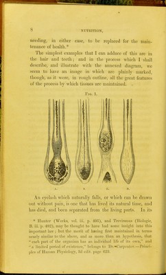
![bulb such an one will be found very different from those that are still living in any period of their age. In the early period of the growth of a dark eyelash, we find its outer end almost uniformly dark, marked only with darker short linear streaks, and exhibiting no distinction of cortical and medullary substance. Not far from its end, however, this distinction is plainly marked; dark as the cortical part may be, the medullary appears like an interior cylinder of much darker granular substance : and in a young hair this con- dition is continued down to its deepest part, where it enlarges to form the bulb. (Fig. 1. a.) Now this enlarge- ment, which is of nearly cup-like form, appears to depend on the accumulation of round and plump nucleated cells, ^vhich, according to their position, are either, by narrowing and elongation, to form the dry fibro-cells of the outer part of the growing and further protruding shaft, or are to be transformed into the air-holding cells of 'the medullary portion. At this time of most active growth, both cells and nuclei contain abundant pigment-matter, and the whole bulb looks nearly black. The sources of the material out of which the cells form themselves are, at least, two; namely, the inner siu-face of the sheath, or capsule, which dips into the skin, enveloping the hair, and the surface of the vascular pulp, which fits in a conical cavity in the bottom of the hair-bulb. Such is the state of parts so long as the growing hair is all dark. But, as it approaches the end of its existence, it seems to give tokens of advancing age, by becoming grey. (Fig. 1. B, 0.) Instead of the almost sudden enlargement at its bulb, the hair only swells a little, and then tapers nearly to a point; the conical cavity in its base is contracted, and hardly domonstrable, and the cells produced on the inner surface of the capsule contain no ])article of pigment. Still, for some time it continues thus to live, and grow, aiul we](https://iiif.wellcomecollection.org/image/b2128913x_0027.jp2/full/800%2C/0/default.jpg)