Volume 4
The cyclopaedia of anatomy and physiology / edited by Robert B. Todd.
- Date:
- 1836-1859
Licence: Public Domain Mark
Credit: The cyclopaedia of anatomy and physiology / edited by Robert B. Todd. Source: Wellcome Collection.
Provider: This material has been provided by King’s College London. The original may be consulted at King’s College London.
114/702 (page 98)
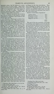
![and rectum, and more rarely about the eyelids, scalp, neck, and prepuce. The matter itself is homogeneous, but differs in consistence in different parts of the cyst — it may be almost fluid in some. Dr. Lever* describes a cyst between the uterus and vagina which gave out a quantity of fluid matter, when punctured, that looked like dripping when cold. In ovarian cysts steatoma is frequently associated with hair. (D.) Cholcstericfats.—Plates of cholesterinf are frequently found in the fluid of hydrocele and of cysts of the thyroid gland. Rayer J found them in a cyst of the kidney, in a sub- ject whose aorta contained several small tu- mours a little above the bifurcation, seated under the lining membrane, and composed in great part of cholesteric scales. § In a female who lately died in our wards with obstructed bowels from stricture of the rectum, a large cyst lying behind the right psoas muscle con- tained cholesterin in atheromatous-looking patches on the inner surface of its wall : here it was undergoing calcification. Cholesterin has been found in scales among pus of an abscess near a carious tooth ||, and of an abscess near an anchylosed joint-lf In such cases it disappears from the secreted matters before suppuration ceases. Cholesterin occurs occasionally in various morbid growths,— for example, in the different varieties of cancer. Closely allied to (if not sometimes identical with) cholesterin in chemical constitution, is a fatty product, for which the name of chole- steatoma has recently been proposed by Miiller. This substance occurs in the forms of (1) tiL- mours ; (2) granules ; (3) patches ; (4) scales. (]). Tumours composed of this material are commonly of the consistence of tallow ; firmer than the brain when found in that organ. They are not lobulated, but frequently mam- millated on the surface; uneven, with a gene- ral tendency to roundness ; surrounded with a capsule of delicate fibrous structure; varying in size from a walnut to the clenched fist and upwards. Of sixteen recorded cases the brain was the seat of the tumour in seven; the bones in three; the utero-rectal cellular tissue in two; subcutaneous cysts in three; a large cysto-sarcoma of the breast in one. The interior of the mass has a shining white and semi-transparent aspect, either generally or in some spots only, while in others the white colour is dull, Miiller found that the substance shrinks and becomes yellowish by desiccation. All who have observed it describe it as com- posed of delicate laminae, for the most part * Med. Chir. Trans, vol. xxiii. t Becquerel and Rodier affirm tliat the proportion of cliolesterin in the blood increases in persons of both sexes from the age of forty to fifty: hence, perhaps, the greater frequency of separation of this fat in old people. % ]Maladies des Reins, t. iii. p, 541. § See also Christison, Ed. Med. and Surg. Jom-n. vol. xxxii. p. 278. II Caventou, Journ. de Pharraacie, t. xi. p. 463. 1825. ^ Nasse, MttUer's Archiv, 1840, Ileft. iii. S. 2G7. arranged concentrically, and easily separated: it is completely extra-vascular. The laminae and the matter interposed be- tween them possess different structures, (a.) The laminai consist of superimposed strata of cells, rendered pentagonal, hexagonal, or polygonal, by lateral mutual pressure, resem- bling, except in size (they are only half as large, averaging a diameter of .00081 of an inch), the cells of sheep's fat {fig. 91) ; easily Pohiqonal cells of cholesteatoma magnified 290 times. (After Muller.) separated from each other, transparent and pale; possessing neither nucleus nor central granules, and admitted to be hollow, rather from the analogy of sheep's fat, than proved to be so by observation. The substance form- ing the cells is distinct in its nature from fat, as it is neither dissolved nor deprived of its laminated appearance by boiling alcohol. (/?>.) The inter-laminar matter consists of crystals tabular and lamellar. The tabular are in greatest abundance ; generally short, broad, and rectangular, but frequently narrow and riband-like, and probably composed of pure cholesterin (which appears under the micro- scope in the form of rhombic tables), as acids and alkalies do not affect them. The lamellar crystals look like aciculae of stearin from their being gathered into bundles ; but when deposited from their aethereal secre- tion they are distinctly lamellar, and pointed at both ends. (2.) Granules. — We agree with Miiller in believing that the pearly looking globules described by Cruveilhier in a cancerous growth of the testicle* were very probably composed of cholesteatomatous matter. (.3.) Patches.—Dupuytren observed patches of this substance on the surface of a urinary fistula; Miiller a stratum of it covering a can- cerous ulcer of the mamma. (4.) Scales.—The fluid of hydrocele and of tumours of the thyroid gland frequently con- tains scales of a pearly looking matter, some- times collected abundantly at the most de- pendent part of the cavity. This matter, commonly believed to consist of cholesterin, is not always so composed. Dr. Bostockf found it essentially different from adipocere and from cholesterin, not soluble in water or in alcohol, but partially so in aether, and in- capable of saponification by potash. Miiller found the alcoholic or aethereal solution of the tuberiform variety yielded no oil on eva- poration, but a fine granular fat, probably stearin, with lanceolate lamellar crystals, * An. Path. livr. v. tab. i. fig. 2. t Med. Chir. Trans, vol. xv. p. 158.](https://iiif.wellcomecollection.org/image/b2130046x_0004_0114.jp2/full/800%2C/0/default.jpg)