Licence: Public Domain Mark
Credit: Researches in embryology. (Second series) / by Martin Barry. Source: Wellcome Collection.
Provider: This material has been provided by the Royal College of Physicians of Edinburgh. The original may be consulted at the Royal College of Physicians of Edinburgh.
23/84 page 325
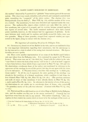
![like nucleus observed by Valentin-I- in globules from various parts of the nervous system. This object was round, colourless, and pellucid, and contained a central dark point, resembling the corpuscle of the above author. The chorion (c/io.) was distinguishable from the fluid /i. Plate VIIL fig. 130, exhibits another of the seven ova crushed. The membrane / is here seen distended and ruptured under the com- pressor. The mulberry-like object—when crushed—not only filled the cavity of that membrane, but a part of it escaped, and its vesicles became pressed together into figures of several sides. The nuclei remained unaltered: their dark central points (nucleoli), however, in this instance had the appearance of globules. In the space between each vesicle and its nucleus, and chiefly around the latter, were now seen granules. Many of those granules, escaped from ruptured vesicles, are repre- sented in the figure, lying in contact with the chorion (cho.). The importance oj examining Ova from the Fallopian Tube. 181. CruikshankJ found ova of the Rabbit in the tube, and we are indebted to him for very important information regarding their minuteness; but the microscope in his day was not in a state to admit of his seeing their internal structure. His figures therefore are mere specks. 182. There exists another representation of an ovum of the Rabbit (and I believe only one) taken from the Fallopian tube. It is contained in a paper by T. Wharton Jones That ovum was one of the third day, found with five others in the tube near where it enters the horn of the uterus, and in size { -^th of an inch) appears to have been between those which I have represented in Plate VI. figs. 109 and 110. My observations corroborate those of the author just mentioned in reference to the appearance of the envelope in ova of this period, but they do not agree with his views as to its real nature. T. Wharton Jonesj] describes this envelope as a thick gela- tinous matter. In all the ova I examined, the outer portion of the envelope was already in the condition of a formed membrane, which condition it had from the first retained (Plate VIII. fig. 128., Plate VI. fig. 110. cho.). (The previous existence and mode of origin of that membrane I have already shown in Plate VI. fig. 104. a. and |3. and in Plate IX. fig. 153.). In reference to the interior of ova of this period, my observations do not enable me to corroborate those of Jones; who remarks^ the granulary matter of the yelk was coherent. (Contrast with Plate VI. fig. 105^ to 110.) 183. Prevost and Dumas-|-|- found no ova of either Dogs or Rabbits in the Fallopian tube, and the smallest ova they saw in the uterus measured Coste:{:| has not figured an ovum from the tube in any animal. This author remarks §§, after con- ception, we have stated, the vesicle which we know to be the analogue of the vesicle of t Ueber den Verlauf und die letzten Enden der Nerven, figs. 51, 52, 70. 1836. X L. c. § Philosophical Transactions, 1837, Part II. plate xvi. fig. 1. II L. c. p. 339. 5[ L. c. p. 339. tt L. c. tl Embryogenie Comparee. §§ pp. 109, 110, V](https://iiif.wellcomecollection.org/image/b2197214x_0025.jp2/full/800%2C/0/default.jpg)