The article Uterus and its appendages from the Cyclopaedia of anatomy and physiology : comprising the normal and abnormal anatomy, physiology and development of the uterus, ovary, parovarium, Fallopian tube, vagina, vulva and placenta / by Arthur Farre.
- Farre, Arthur, 1811-1887.
- Date:
- [1858]
Licence: Public Domain Mark
Credit: The article Uterus and its appendages from the Cyclopaedia of anatomy and physiology : comprising the normal and abnormal anatomy, physiology and development of the uterus, ovary, parovarium, Fallopian tube, vagina, vulva and placenta / by Arthur Farre. Source: Wellcome Collection.
Provider: This material has been provided by The Royal College of Surgeons of England. The original may be consulted at The Royal College of Surgeons of England.
37/182 (page 581)
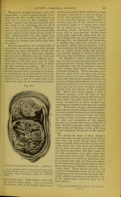
![The growth of these secondary cysts with broad bases, of which a good example is ex- hibited in Jig. 394., is often very irregular, so that one or more of them enlarging with greater rapidity than the rest, encroach upon the cavity of the containing cyst, and fill it more or less completely. This rapid enlarge- ment of the secondary cysts also occasionally causes rupture of their walls and the escape of their contained fluids into the parent cyst, followed by the unrepressed growth of the secondary or tertiary cysts which arise from its surface. After the appearance of a tertiary order of cyst within the secondary ones, their growth occasions so much disturbance of the even outlines of the walls in which they originate, that it is often difficult to trace the order and manner of enlargement of the different series. Nevertheless, with care, these may be often made out even in the complex forms, of which Jig. 395. furnishes an example. Here is re- presented a small portion only of an enor- mously enlarged ovary, consisting of a primary or principal sac, the greater part of which has been cut away, so as to leave a part of its walls visible at a, a, a, and of a more solid basis which was made up of numerous secondary Fig. 395. Compound orproliferus ovarian cyst. ^Ad Nat.) a,a,a, divideil walls of the principal single cyst; b, small simple cyst; c, c, two masses of compound secondary cysts, containing many of a tertiarv order. and tertiary cysts. Both of these orders may be traced m this example. At a, n, a are seen the divided walls of the original parent cyst, whilst springing from these walla at h is a single secondary cyst, and at c, c are two groups of similar cysts aggregated in masses. Tlie lat- ter are, however, examples of compound se- condary cysts, for in the interior of each is contained a series of a tertiary order, which are so numerous as to fill completely the secondary sacculi. By Lebert it is deemed to be still an open question whether these cysts, which apparently spring from the in- terior of the main sac, as represented in Jigs. 394. and 395., are altogether new formations, or whether they were not originally in part developed, and by force of pressure, arising from contiguity of situation, have penetrated and at length grown within the principal cyst, into the interior of which, in this view, therefore, they would form a species of hernia. Dr. Hodgkin *, whose elaborate descrip- tions of ovarian cysts has made known all the principal varieties of form which they assume, distinguishes from the broad-based cysts, al- ready noticed, those which arise by narrow or slender peduncles. These sometimes grow from the walls of the principal cyst, and, in- deed, in almost all cases which I have exa- mined, after the sac has attained a certain size, patches of these pedunculated sacculi may be observed scattered over the interior in various places, but they are more constantly observed growing from the interior of the secondary cyst. These little sacculi appear at first in scattered patches, under the form of little round grains, thickly covering the lining mem- brane which they raise above them, and so closely set that two or three hundred may sometimes be counted in the space of a square inch. When these elongate, mutual pressure causes them to assume a filamentous condition ; but when greater freedom of growth is enjoyed, their extremities commonly dilate into little pouches, or buds of another order S()rout from the sides and extremities of the original growths, and convert them into a multitude of little dendritic processes which roughen the inner surface of the larger cysts, or fill more or less completely the cavities of the smaller ones. If a section be made of these dendritic processes, they are seen usually to be solid at their base, the white fibrous tissue of the parent cyst wall, from which they spring, being easily traced into their stems and branches. But at their extremities they become dilated into little pouches filled with fluid, similar to the little pediculated cysts, with which they are abundantly intermixed. These little cysts and processes are covered by epithelium, and it is probable that they are the active agents in the elimination of the various fluids by which the ovarian cysts, of whatever order, are com- monly filled. These minute processes and vesicles, so abundantly found on the walls of endogenous cysts, are represented in fig. 396., which exhibits a portion of a proliferous cyst of the natural size, covered by them on 'its inner surface. * Loc. citat. and Med. Chir. Trnn.s. Vol. xv. ])(. ii.](https://iiif.wellcomecollection.org/image/b22287449_0039.jp2/full/800%2C/0/default.jpg)