The article Uterus and its appendages from the Cyclopaedia of anatomy and physiology : comprising the normal and abnormal anatomy, physiology and development of the uterus, ovary, parovarium, Fallopian tube, vagina, vulva and placenta / by Arthur Farre.
- Farre, Arthur, 1811-1887.
- Date:
- [1858]
Licence: Public Domain Mark
Credit: The article Uterus and its appendages from the Cyclopaedia of anatomy and physiology : comprising the normal and abnormal anatomy, physiology and development of the uterus, ovary, parovarium, Fallopian tube, vagina, vulva and placenta / by Arthur Farre. Source: Wellcome Collection.
Provider: This material has been provided by The Royal College of Surgeons of England. The original may be consulted at The Royal College of Surgeons of England.
5/182 (page 549)
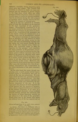
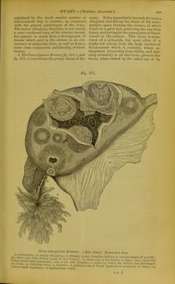
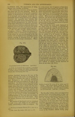
![explained by tlie much smaller number of blooil-vessels that it contains, as compared with the general parenchyma of the ovary. The tunica albuginea, therefore, is not merely a more condensed form of the ovarian stroma, but appears to result from a development of tissues which exist in the stroma in an ele- mentary or embryonic form, as well as from a more close conjunction and blending of those tissues. 2. ThcPurowhi/mnorSlruma,{fig- 37].c,and fig. 372. s) constitutes tlie proper tissue of the ovary. It lies immediately beneath the tunica albuginea, and fdls up the whole of the inter- mediate space between the ovisacs, to which it acts as a germ bed, protecting the ova from injury, and serving for tlie conveyance of blood- vessels to the ovisacs. This tissue is some- times of a pale-pink, but more often of a bright-red colour, from the large niunber of blood-vessels which it contains, whose ar- rangement proceeding from within, and radi- ating outwardly in all directions, gives to this tissue, when viewed by the naked e3'e or by Fig. 371, Ovary enlarged four diameters. {After Coste.) Dissected to shew,](https://iiif.wellcomecollection.org/image/b22287449_0007.jp2/full/800%2C/0/default.jpg)