The article Uterus and its appendages from the Cyclopaedia of anatomy and physiology : comprising the normal and abnormal anatomy, physiology and development of the uterus, ovary, parovarium, Fallopian tube, vagina, vulva and placenta / by Arthur Farre.
- Farre, Arthur, 1811-1887.
- Date:
- [1858]
Licence: Public Domain Mark
Credit: The article Uterus and its appendages from the Cyclopaedia of anatomy and physiology : comprising the normal and abnormal anatomy, physiology and development of the uterus, ovary, parovarium, Fallopian tube, vagina, vulva and placenta / by Arthur Farre. Source: Wellcome Collection.
Provider: This material has been provided by The Royal College of Surgeons of England. The original may be consulted at The Royal College of Surgeons of England.
6/182 (page 550)
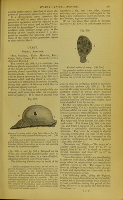
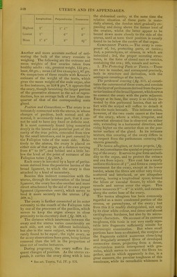
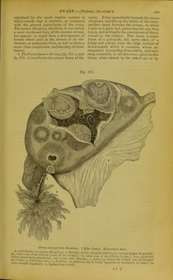
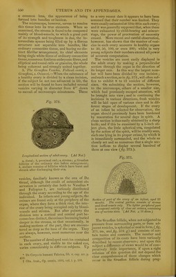
![a common lens, the appearance of being formed into bundles or laminae. The microscope, however, serves to resolve this tissue into its true elements. When so examined, the stroma is found to be composed mainly of blood-vessels, to which a great part of its strength and toughness is due, the in- termediate spaces being filled up by a fibrous structure not separable into bundles, like ordinary connective tissue, and having no dis- tinct fibrillar arrangement, its chief elements being single white fibres of ordinary connective tissue.numerous fusiform embryonic fibres,and elliptical and round cells or granules, the whole being coherent and strongly united together. 3. The Graafian Vesicles. Folliculi ovarii, s. Graafiani, s. Ovisacci.—When the substance of a healthy ovary is divided by a clean incision, if the subject be not too advanced in life, the section will be found to have included several vesicles varying in diameter from 4' down to sacculi of microscopic minuteness. These Fig. 372. Longitudinal section of adult ovary. {Ad Nat.) a, distal; b, proximal end; s, stroma; g, GraaOan follicles of the ordinary size before enlargement; h, stellate remains of follicles which have burst and shrunk after discliarging their ova. vesicles, familiarly known as the ova of De Graaf, although the credit of antecedent ob- servation is certainly due both to Vesalius * and Fallopius f, are variously distributed through the ovary according to the age of the individual. In infants and young subjects, the ovisacs are found only at the periphery of the organ, where they form a thick rind, the inte- riol- of the ovary being occupied only by blood- vessels and stroma. But after puberty the division into a cortical and central part be- comes less distinct, theovisacs becoming buried deeper in the stroma, so that occasionally, in making sections of the part, they are encoun- tered as deep as the base of the organ. They are always, however, most numerous near the surface. The number of developed vesicles contained in each ovary, and visible to the naked eye, varies considerably in different subjects. Up * De Corporis humani Fabrica, lib. v. cap. xv. p. 459. t Obs. Anat., Op. omniii, 160G, vol. i. p. lOG. to a very recent date it appears to have been assumed that their number was limited. They wereusually estimated at 12 to 20 in each ovary; and it was generally supposed that, when these were exhausted by child-bearing and miscar- riage, the power of procreation of necessity ceased. More recent and careful observation, however, has shown that the number of vesi- cles in each ovary amounts in healthy organs to 30, 50, 100, or even 200; whilst in very young subjects their numbers exceed all power of accurate computation. The vesicles are most easily displayed in the adult ovary by making a perpendicular section through the organ in the direction of its longer axis. In this way the largest num- ber will have been divided by one incision ; and such a section, as in _/?g. 372., will often suf- fice to exhibit 8 to 12 vesicles of different sizes. On submitting the section, however, to the microscope, others of a smaller size, which had previously escaped attention, will be brought into view ; and in continuing the incisions in various directions, fresh vesicles will be laid open of various sizes and in dif- ferent stages of development. If the ovary of an infant be selected for observation, the organ should previously have been hardened by maceration for several days in spirit. A clean section is thus easily obtained by a sharp knife; and if this be examined by a 1-inch ob- ject glass, the little s|)herical ova, coagulated by the action of the spirit, will be readily seen, each one lying in its proper ovisac, by which it is immediately surrounded, and the whole so closely set and so numerous that a single sec- tion suffices to display several hundred of them at one view (Jig. 373.). Fig. 373. Section of ])art of the ovary of an infant, aged 20 months. The central portion co7isists of stroma and blood-vessels only. The lighter peripheral part is composed entirely of close-set ovisacs, containing ova of various sizes. (Ad Nat. x 16 diam.) The Graafian follicle, when not subjected to pressure from surrounding parts, or from ad- jacent vesicles, is spherical or oval in form, (fig. 371. DD, and fig. 372. g) and consists of cer- tain tunics and contents. The number and composition of its coats have been variously described by recent observers ; and upon this subject a difference of views would be of com- paratively little importance, if upon a right solution of this question did not depend the clear comprehension of those changes whicii occur in the (fraafian follicle during preg-](https://iiif.wellcomecollection.org/image/b22287449_0008.jp2/full/800%2C/0/default.jpg)