The article Uterus and its appendages from the Cyclopaedia of anatomy and physiology : comprising the normal and abnormal anatomy, physiology and development of the uterus, ovary, parovarium, Fallopian tube, vagina, vulva and placenta / by Arthur Farre.
- Farre, Arthur, 1811-1887.
- Date:
- [1858]
Licence: Public Domain Mark
Credit: The article Uterus and its appendages from the Cyclopaedia of anatomy and physiology : comprising the normal and abnormal anatomy, physiology and development of the uterus, ovary, parovarium, Fallopian tube, vagina, vulva and placenta / by Arthur Farre. Source: Wellcome Collection.
Provider: This material has been provided by The Royal College of Surgeons of England. The original may be consulted at The Royal College of Surgeons of England.
7/182 (page 551)
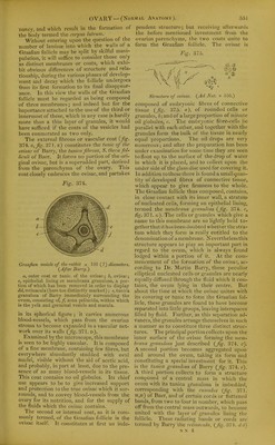
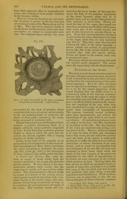
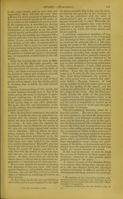
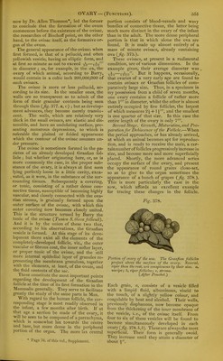
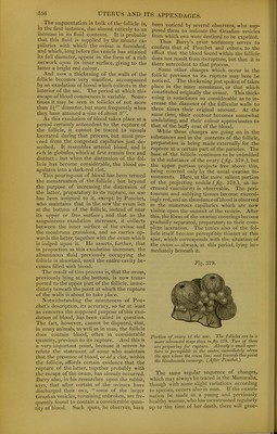
![nancy, and which result in the body termed tlie corpus luteum. Without entering upon the question of the number of laminae into which the walls of a Graafian follicle mav be split by skilful mani- pulation, it will suffice to consider those only as distinct membranes or coats, which exhi- bit obvious differences of structure and rela- tionship, during the various ])hases of develop- ment and decay which the follicle undergoes from its first formation to its final disappear- ance. In this view the walls of the Graafian follicle must be regarded as being composed of three membranes; and indeed but for the importance attached to the use of the third or innermost of these, which in any case is hardly more than a thin layer of granules, it would have sufficed if the coats of the vesicles had been enumerated as two only. The external fibrous or vascular coat {fg. 374. a, jig. 371, e) constitutes the tunic of the ovisac of Barry, the tunica fibrosa, S. theca fol- licuH of Baer. It forms no portion of the ori- ginal ovisac, but is a superadded part, derived from the parenchyma of the ovary. This coat closely embraces the ovisac, and partakes Fig. 374. OVARY — (Normal Anatomy). the formation of pendent structure 551 Graafian vesicle of the rabbit x 100 ( ?) diameters. (^Afler Barry.) a, outer coat or tunic of the ovisac; b, ovisac; c, epithelial lining or membrana granulosa, a por- tion of which has been removed in order to display dd, retinacula (here too distinctly marked) ; e, tunica granulosa of Barry immediately surrounding the ovum, consisting of,/, zona pellucida, within which is the yelk and germinal vesicle and macula. in its spherical figure ; it carries numerous blood-vessels, which pass from the ovarian stroma to become expanded in a vascular net- work over its walls {fig. 371. d). Examined by the microscope, this membrane is seen to be highly vascular. It is composed of a fine membrane, containing few fibres, but everywhere abundantly studded with oval nuclei, visible without the aid of acetic acid, and probably, in part at least, due to the pre- sence of so many blood-vessels in its tissue. This coat contains no oil globules. Its chief use appears to be to give increased support and protection to the true ovisac which it sur- rounds, and to convey blood-vessels from the ovary for its nutrition, and for the supply of the fluids which the ovisac contains. The second or internal coat, as it is com- monly termed, of the Graafian fi)llicle is the ovisac itself. It constitutes at first an inde- but receiving afterwards the before mentioned investment from the ovarian parenchyma, the two coats unite to form the Graafian follicle. The ovisac is Structure of ovisac. {Ad Nat. x 350.) composed of embryonic fibres of connective tissue {fig. 375. a), of rounded cells or granules, 6; and of a large proportion of minute oil globules, c. The embryonic fibre-cells lie parallel with each other, and together with the granules form the bulk of the tissue in nearly equal proportions. The oil drops are very numerous; and after the preparation has been under examination for some time they are seen to float up to the surface of the drop of water in which it is placed, and to collect upon the under side of the glass disc used for covering it. In addition to these there is found a small quan- tity of developed fibres of connective tissue, which appear to give firmness to the whole. The Graafian follicle thus composed, contains, in close contact with its inner wall, a stratum of nucleated cells, forming an epithelial lining, termed the membrana granulosa {fig. 374-. c, fig. 371. g). The cells or gi'anules which give a name to this membrane are so lightly held to- gether that it has been doubted whetlier the stra- tum which they form is really entitled to the denomination of amembrane. JSTeverthelessthis structure a|)pears to play an important part in regard to the ovum, which is always found lodged within a portion of it. At the com- mencement of the formation of the ovisa':, ac- cording to Dr. Martin Barry, these peculiar elliptical nucleated cells or granules are nearly equally diffused through the fluid which it con- tains, the ovum lying in their centre. But about the time at which the ovisac unites witii its covering or tunic to form the Graafian fol- licle, these granules are found to have become separated into little groups, leaving interspaces filled by fluid. Further, as this separation ad- vances, the granules arrange themselves in such a manner as to constitute three distinct struc- tures. The principal portion collects upon the inner surface of the ovisac forming the vicm- brana granulosa just described {fg. 374. c). A second portion becomes aggregated upon and around the ovum, taking its form and constituting a special investment for it. This is the tunica granulosa of Bai'ry {fig. 374. e). A third portion collects to form a structure composed of a central mass in which the ovum with its tunica aranulosa is imbedded. 371. corresponding with the cumulus H,ii) of Baer, and of certain conls orflattened bands, from two to four in number, which pass off from the central mass outwards, to become united with the layer of granules lining the follicle. These radiating bauds or cords arc termed by Barry the rcLinaculti, {fig. 371. d d)](https://iiif.wellcomecollection.org/image/b22287449_0009.jp2/full/800%2C/0/default.jpg)