Hand atlas of human anatomy / by Werner Spalteholz ; translated from the 3rd German edition by Lewellys F. Barker.
- Spalteholz, Werner, 1861-1940. Handatlas der Anatomie des Menschen. English
- Date:
- 1900-
Licence: In copyright
Credit: Hand atlas of human anatomy / by Werner Spalteholz ; translated from the 3rd German edition by Lewellys F. Barker. Source: Wellcome Collection.
31/896 (page 19)
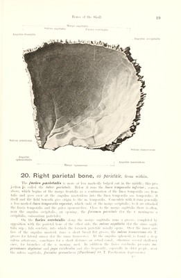
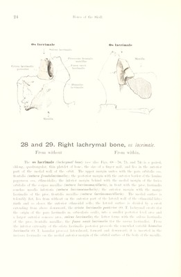
![I Angulus frontalis Margo Sulcus sagittalis sagittal is I* acics cerebralis Sulcus transvcrsns Sulcus arteriosus Angulus occipitalis Angu lus spheuoidalis Margo s(jnainosus Angulus mastoideus 20. Right parietal bone, os parietale, from within. The facies parietalis is more or less markedly bulged out in the middle; this pro¬ jection [is called the tuber parietale. Below it runs the tinea temporalis inferior, convex above, which begins at the margo frontalis as a continuation of the linea temporalis oss. fron¬ talis and goes over at the angulus mastoideus into the linea temporalis oss. temporalis; it itself and the field beneath give origin to the m. temporalis. Concentric with it runs generally a less marked linea temporalis superior, which ends at the margo occipitalis; to it are attached the fascia temporalis and the galea aponeurotica. Close to the margo sagittalis there is often, near tlx* angulus occipitalis, an opening, the foramen parietale (for the r. meningeus a occipitalis, emissarium parietale). On tlx* facies cerebralis along the margo sagittalis runs a groove, completed bv apposition with the parietal hone of the other side, the sulcus sagittalis (for the sinus sagit¬ talis sup.; falx cerebri), into which the foramen parietale usually opens. Over the inner sur¬ face of the angulus mastoid, runs a short broad Hat groove, the sulcus transccrsus (0. T. groove for lateral sinus) (for the sinus transfers.). At the angulus sphenoid, is found a deep sulcus arteriosus, sometimes for a short distance an actual canal, otherwise several shallower ones, for branches of the a. meiiing. med. In addition the facies cerebralis presents im- pressioues digitatae and jug a cerebralia and also frequently, especially in older people, near the sulcus sagittalis, foveolac granulares [Pacchioni] (0. T. Pacchionian depressions).](https://iiif.wellcomecollection.org/image/b29336211_0031.jp2/full/800%2C/0/default.jpg)