Report of the Pellagra Commission of the State of Illinois, November, 1911.
- Illinois. Pellagra Commission.
- Date:
- 1912
Licence: In copyright
Credit: Report of the Pellagra Commission of the State of Illinois, November, 1911. Source: Wellcome Collection.
Provider: This material has been provided by Royal College of Physicians, London. The original may be consulted at Royal College of Physicians, London.
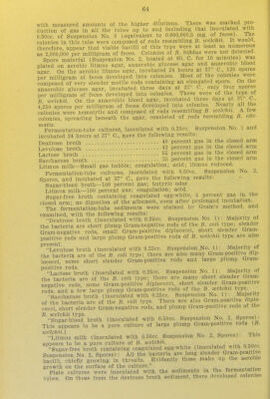
69/279 page 61
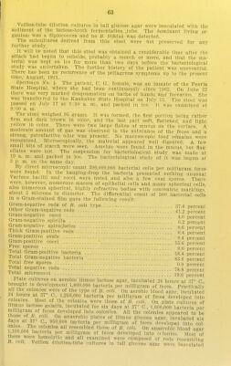
![Saccharose broth (0.25cc. Suspension No. 1): Majority of the bacteria are Gram-positive diplococci of medium size, many Gram-negative rods re- sembling B. coll, and many Jong, thick Gram-positive rods are also present. Litmus milk (0.25cc. Suspension No. 1): Majority of the bacteria are Gram-positive diplococci; rods of B. coH and B. toelchii types are also present. Dextrose broth (O.SOcc. Suspension No. 5): Majority of the bacteria are Gram-positive diplococci of medium siz.e. Numerous rods resembling B. coli and a few thick Gram-positive rods are also present. Levulose broth (O.SOcc. Suspension No. 5) and lactose broth (O.SOcc. Suspension No. S): Same as dextrose broth. Saccharose broth (O.SOcc. Suspension No. 5): Great majority of the bacteria are short plump Gram-negative rods of B. coli type; very few Gram- positive diplococci. Litmus milk (O.SOcc. Suspension No. 5): Majority of the bacteria are Gram-positive diplococci of medium size. Short plump Gram-positive rods (B. welchii) and small Gram-positive cocci are also present. Sugar-blood broth (O.SOcc. Suspension No. 2, spores): Majority of the bacteria are plump Gram-positive rods; others are decolorized, but may be of the same species (B. welchii?). Litmus milk (O.SOcc. Suspension No. 2, spores): Apparently a pure culture of B. ivelcMi. Sugar-free broth containing coagulated egg-white: Slender Gram-positive rods in threads; a few large, plump Gram-negative rods. Culture strains derived from this stool were not preserved. It will be noted that this was diarrhea] stool passed naturally several weeks after the patient had begun to recover from the attack of pellagra. The desquamation of the pigmented- epithelium was completed soon afterward, and there has been no further sign of pellagra up to the present time August, 1911. Specimen No. 3. The patient, M. R., female, was an inmate of the Peoria State Hospital, where she had been continuously for four years. She had an attack of pellagra in 1909. On June 22, 1910, she had a dark chocolate- brown discoloration on the backs of hands and forearms, but no other mani- festations of pellagra. She was transferred to the Kankakee State Hospital on July 13, 1910. The stool examined was passed naturally at 2:40 a. m. on July 17th, and was packed in ice. It was examined at 8:30 a. m. on the same day. The stool was very soft, almost mushy in consistency, with a light pink- ish brown color and a normal odor. Macroscopic food remnants were not found. Microscopic examination showed some bits of broken starch, but no meat residue. On the whole the food appeared to be well digested. No protozoa were detected. The suspension for bacteriological study was pre- pared from the mixed feces and was packed in ice at 9:00 a. m. where it remained until July 19th, at 9:3S a. m., when the bacteriological study was By the microscopic counting method 180,000,000 bacterial cells per milli- gram of feces were found. Study of the hanging-drop preparation showed very few spores and no spirochetes. Differential count of 500 cells, stained by Gram's method, gave the following results: begun. Gram-negative reds of B. coli type Other Gram-negative rods Gram-negative cocci 46.0 percent 29.0 percent 0.0 percent 0.4 percent 0.6 percent 3.2 percent 0.8 percent SO.O percent 0.0 percent 24.6 percent 75.4 percent Gram-negative spirilla Thick Gram-positive rods Slender Gram-positive rods . Oval Gram-positive bacilli .. Gram-positive cocci Spores Total Gram-positive bacteria Total Gram-negative bacteria](https://iiif.wellcomecollection.org/image/b2398322x_0069.jp2/full/800%2C/0/default.jpg)