Report of the Pellagra Commission of the State of Illinois, November, 1911.
- Illinois. Pellagra Commission.
- Date:
- 1912
Licence: In copyright
Credit: Report of the Pellagra Commission of the State of Illinois, November, 1911. Source: Wellcome Collection.
Provider: This material has been provided by Royal College of Physicians, London. The original may be consulted at Royal College of Physicians, London.
71/279 page 63
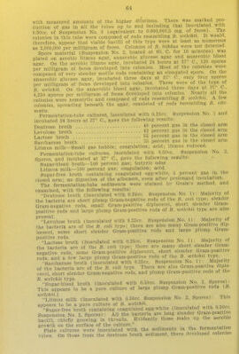
![Veillon-tube dilution .cultures in tall glucose agar were inoculated with the sediment of the lactose-broth fermentation .tube. The dominant living or- ganism was a diplococcus and no B. Mfidu's was detected. The subcultures derived from this stool were not preserved for any further study. It will be noted that this stool was obtained a considerable time after the attack had begun to subside, probably a month or more, and that the ma- terial was kept on ice for more than two days before the bacteriological study was undertaken. The further history of the patient was uneventful. There has been no recurrence of the pellagrous symptoms up to the present time, August, 1911. Specimen No. J,. The patient, C. G., female, was an inmate of the Peoria State Hospital, where she had been continuously since 1902. On .June 22 there was very marked desquamation on backs of hands and forearms. She was transferred to the Kankakee State Hospital on July 13. The stool was passed on July 17 at 5:30 a. m., and packed in ice. It was examined at 9:00 a. m. The stool weighed 95 grams. It was formed, the first portion being rather firm and dark brown in color, and the last part soft, flattened, and light yellow in color. There were two large flakes of mucus on the surface. A moderate amount of gas was observed in the substance of the feces and a strong, putrefactive odor was present. No macroscopic food remains were recognized. Microscopically, the material appeared well digested. A few small bits of starch were seen. Amebae were found in the mucus, but flag- ellates were not. The suspension for bacteriological study was made at 10 a. m. and packed in ice. The bacteriological study of it was begun at 3 p. m. on the same day. By direct microscopic count 300,000,000 bacterial cells per milligram feces were found. In the hanging-drop the bacteria presented nothing unusual Various bacilli and cocci were noted and also a few oval spores There were, however, numerous masses of epithelial cells and many spherical cells, also numerous spherical, highly refractive bodies with concentric markings about 5 microns in diameter. The differential count of 500 bacterial cells m a Gram-stained fllm gave the following result: Gram-negative rods of B. coli type - 37 6 percent Other Gram-negative rods 412 percent Gram-negative cocci [ ' ' 4 0 percent Gram-negative spirilla .. 0 2 percent Gram-negative spirochetes 0 6 percent Thick Gram-positive rods \]] o.4 percent Gram-positive ovals 0 4 percent Gram-positive cocci • IS.'g percent Free spores 0 0 percent Total Gram-positive bacteria 16 4 percent Total Gram-negative bacteria 83.6 percent Tota free spores 0.0 percent Tota negative, rods 78.8 percent Total micrococci 19 6 p^j.^^nt Plate cultures on aerobic litmus lactose agar, incubated 24 hours at 37° C brought to development 1,400,000 bacteria per milligram of feces. Practically all the colonies were of the type of B. coli. On aerobic blood agar, incubated 24 hours at 37° C, 1,250,000 bacteria per milligram of feces developed into colonies. Most of the colonies were those of B. coli. On plate cultures of litmus lactose gelatin, incubated for six days at 37° C, 1,600,000 bacteria per milligram of feces developed into colonies. All the colonies appeared to be those of B coli. On anaerobic plates of litmus glucose agar, incubated six Clays at 37 C, 950,000 bacteria per milligram of feces developed into col- ?°innnnJ v,^ Colonies all resembled those of B. coli. On anaerobic blood agar 1,100,000 bacteria per milligram of feces developed into c( lonies Most of these were hemolytic and all examined were composed of rods resembling B. colt. Veillon dilution-tube cultures in tall glucose agar were inoculated](https://iiif.wellcomecollection.org/image/b2398322x_0071.jp2/full/800%2C/0/default.jpg)