Licence: Public Domain Mark
Credit: The pathology of the membrane of the larynx and bronchia. Source: Wellcome Collection.
Provider: This material has been provided by the Harvey Cushing/John Hay Whitney Medical Library at Yale University, through the Medical Heritage Library. The original may be consulted at the Harvey Cushing/John Hay Whitney Medical Library at Yale University.
48/232 page 42
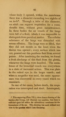
![whose body I opened, within the membrane there was a diameter exceeding two eighths of an inch*. Through a tube of this diameter, an adult can support respiration for a consi- derable time, without great inconvenience. In these bodies the air vessels of the lungs were full of a fluid, which it was impossible to distinguish from purulent matter. The cellular substance of the lungs was distended with serous effusion. The lungs were so filled, that they did not recede in the least when the thorax was opened ; every surface which was cut, poured out this puriform fluid from several points. After insulating the trachea, we found a fresh discharge of this fluid from the glottis, whenever the lungs were handled. The mem- brane of the bronchia] vessels was everywhere in a state of increased action. This was plain to the naked eye in the larger vessels; and when a magnifier was used, the same appear- ance was discovered in every vessel which we examined. In one of the cases, before death, the respi- ration was interrupted and short. Interrupted, * This eDgraving (Plate VII.), taken from a drawing of the trachea after Croup, will illustrate my assertion, that there is sufficient space left within the adventitious membrane for the transmission of the air. This drawing was made without any view to the question which 1 am discussing,](https://iiif.wellcomecollection.org/image/b21030352_0048.jp2/full/800%2C/0/default.jpg)


 No text description is available for this image
 No text description is available for this image
 No text description is available for this image