Practical anatomy : a manual of dissections / by Christopher Heath.
- Christopher Heath
- Date:
- 1881
Licence: Public Domain Mark
Credit: Practical anatomy : a manual of dissections / by Christopher Heath. Source: Wellcome Collection.
Provider: This material has been provided by The University of Glasgow Library. The original may be consulted at The University of Glasgow Library.
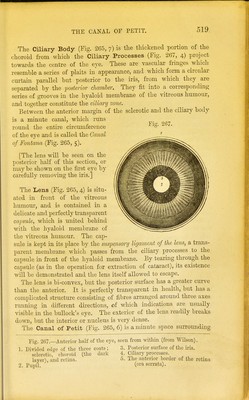
609/646 (page 519)
![THE CANAL OF PETIT. The Ciliary Body (Fig. 265, 7) is the thickened portion of the choroid from which the Ciliary Processes (Fig. 267, 4) project towards the centre of the eye. These are vascular fringes which resemble a series of plaits in appearance, and which form a circular curtain parallel but posterior to the iris, from which they are separated by the -posterior chamber. They fit into a corresponding series of grooves in the hyaloid membrane of the vitreous humour, and together constitute the ciliary zone. Between the anterior margin of the sclerotic and the ciliary body is a minute canal, which runs round the entire circumference of the eye and is called the Canal of Fontanel (Fig. 265, 5). [The lens will be seen on the posterior half of this section, or may be shown on the first eye by carefully removing the iris.] The Lens (Fig. 265, 4) is situ- ated in front of the vitreous humour, and is contained in a delicate and perfectly transparent capsule, which is united behind with the hyaloid membrane of the vitreous humour. The cap- sule is kept in its place by the suspensory ligament of the lens, a trans- parent membrane which passes from the ciliary processes to the capsule in front of the hyaloid membrane. By tearing through the capsule (as in the operation for extraction of cataract), its existence will be demonstrated and the lens itself allowed to escape. The lens is bi-convex, but the posterior surface has a greater curve than the anterior. It is perfectly transparent in health, but lias a complicated structure consisting of fibres arranged around three axes running in different directions, of which indications are usually visible in the bullock's eye. The exterior of the lens readily breaks down, but the interior or nucleus is very dense. The Canal of Petit (Fig. 265, 6) is a minute space surrounding Eig. 267.—Anterior half of the eye, seen from within (from Wilson). 1 Divided ed<*o of the three coats; 3. Posterior surface of the iris. sclerotic,3 choroid (the dark 4. Ciliary processes. layer), and retina. 5. The anterior border of the retina 2. Pupil. (ora serrata).](https://iiif.wellcomecollection.org/image/b2145887x_0609.jp2/full/800%2C/0/default.jpg)