Practical anatomy : a manual of dissections / by Christopher Heath.
- Christopher Heath
- Date:
- 1881
Licence: Public Domain Mark
Credit: Practical anatomy : a manual of dissections / by Christopher Heath. Source: Wellcome Collection.
Provider: This material has been provided by The University of Glasgow Library. The original may be consulted at The University of Glasgow Library.
610/646 (page 520)
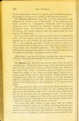
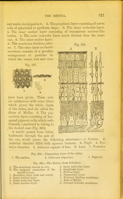
![the lens immediately around the capsule, and is bounded in front by the suspensory ligament and behind by the hyaloid membrane. The Vitreous Humour (Fig. 265, 13) is the transparent body filling all the posterior part of the eyeball. It is a gelatinous sub- stance contained in a transparent membrane called the hyaloid membrane, and is traversed by, numerous delicate and perfectly transparent septa. The existence of these may be demonstrated by crushing the vitreous humour with the fingers, when the fluid portion will drain away. In the eye from which the vitreous humour has been removed the retina will fall together, leaving the inner surface of the choroid exposed, and in the bullock's eye the tapetum lucidum will be seen. This is the coloured appearance which is peculiar to the lower animals, and is due to the presence of a thick layer of wavy fibrous tissue outside the choroidal epithelium. The object of this is to reflect the rays through the retina a second time, and thus to enable the animal to see with a very small amount of light. [The retina is best seen by looking through the vitreous humour of an eye from which the iris and lens have been removed.] The Retina (Fig. 265, 9) is the nervous coat of the eye and its essential part. It lines the choroid coat, being connected posteriorly with the optic nerve, and extends in front nearly as far forward as the ciliary body, where it terminates about one-eighth of an inch behind the margin of the cornea in.a finely jagged border, the ora serrata, being thicker behind than in front. During life the retina is transparent, so that the vascular choroid can be seen through it with the ophthalmoscope, but after death it has a greyish colour and is thrown into folds, owing to the diminution of the tension of the globe. The entrance of the optic nerve may be seen about one-tenth of an inch to the inner side of the axis of the eye, and radiating from this may be seen the branches of the arteria centralis retina;, which enters the eye at this point, and its accompanying veins. In the axis of the eyeball in the human eye is the yellow spot of Soemmering, the most sensitive point, which consists of a small elevation, in the centre of which is a minute depression, the fovea centralis. The retina (Fig. 269) consists of eight layers with two delicate limiting membranes, which are as follows, beginning from the inner surface :—a. The membrana limitans interna. 1. The fibrous layer continuous with the optic nerve (the fibrillte of which here lose their white substance of Schwann), and having numerous ganglionic cells](https://iiif.wellcomecollection.org/image/b2145887x_0610.jp2/full/800%2C/0/default.jpg)