On the myology of the sciuromorphine and hystricomorphine rodents / by F.G. Parsons.
- Frederick Gymer Parsons
- Date:
- 1894
Licence: Public Domain Mark
Credit: On the myology of the sciuromorphine and hystricomorphine rodents / by F.G. Parsons. Source: Wellcome Collection.
Provider: This material has been provided by The Royal College of Surgeons of England. The original may be consulted at The Royal College of Surgeons of England.
35/48 page 283
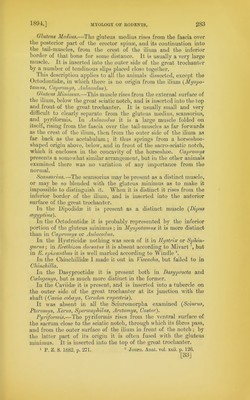
![Gluteus Medius.—The gluteus medius rises from the fascia over the posterior part of the erector spin®, and its continuation into the tail-muscles, from the crest of the ilium and the inferior border of that hone for some distance. It is usually a very large muscle. It is inserted into the outer side of the great trochanter by a number of tendinous slips placed close together. This description applies to all the animals dissected, except the Octodontidae, in which there is no origin from the ilium (Myopo- tamus, Capromys, Aulacodus). Gluteus Minimus.—This muscle rises from the external surface of the ilium, below the great sciatic notch, and is inserted into the top and front of the great trochanter. It is usually small and very dillicult to clearly separate from the gluteus medius, scansorius, and pyriformis. In Aulacodus it is a large muscle folded on itself, rising from the fascia over the tail-muscles as far forwards as the crest of the ilium, then from the outer side of the ilium as far back as the acetabulum ; it thus springs from a horseshoe- shaped origin above, below, and in front of the sacro-sciatic notch, which it encloses in the concavity of the horseshoe. Capromys presents a somewhat similar arrangement, but in the other animals examined there was no variation of any importance from the normal. Scansorius.—The scansorius may be present as a distinct muscle, or may be so blended with the gluteus minimus as to make it impossible to distinguish it. When it is distinct it rises from the inferior border of the ilium, and is inserted into the anterior surface of the great trochanter. In the Dipodidae it is present as a distinct muscle (Dipus cegyptius). In the Octodontidae it is probably represented by the inferior portion of the gluteus minimus ; in Myopotamus it is more distinct than in Capromys or Aulacodus. In the Ifvstricidae nothing was seen of it in TTystrix or Spliin- gurus; in Erethizon dorsatus it is absent according to Mivart but in E. epixantlius it is well marked according to Windie 2. In the Chiuchillidae 1 made it out in Viscacha, but failed to in Chinchilla. In the Dasyproctidae it is present both in Dasyprocta and Coelogenys, but is much more distinct in the former. In the Caviidae it is present, and is inserted into a tubercle on the outer side of the great trochanter at its junction with the shaft (Cavia cobaya, Ceredon rupestris). It was absent in all the Sciuromorpha examined (Sciurus, Pteromys, Xerus, Spermophilus, Arctomys, Castor). Pyriformis.—The pyriformis rises from the ventral surface of the sacrum close to the sciatic notch, through which its fibres pass, and from the outer surface of the ilium in front of the notch; by the latter part of its origin it is often fused with the gluteus minimus. It is inserted into the top of the great trochanter. 1 P. Z. S. 1882, p. 271- 2 Journ. Anat. vol. xxii. p. 126. [33]](https://iiif.wellcomecollection.org/image/b2238635x_0037.jp2/full/800%2C/0/default.jpg)