Nouveaux élements de médecine opératoire : accompagnés d'un atlas de 20 planches in-4o gravées, représentant les principaux procédés opératoires et un grand nombre d'instruments de chirurgie / par Alf. A.L.M. Velpeau.
- Velpeau, A. (Alfred Armand Louis Marie), 1795-1867.
- Date:
- 1832
Licence: Public Domain Mark
Credit: Nouveaux élements de médecine opératoire : accompagnés d'un atlas de 20 planches in-4o gravées, représentant les principaux procédés opératoires et un grand nombre d'instruments de chirurgie / par Alf. A.L.M. Velpeau. Source: Wellcome Collection.
Provider: This material has been provided by King’s College London. The original may be consulted at King’s College London.
13/102
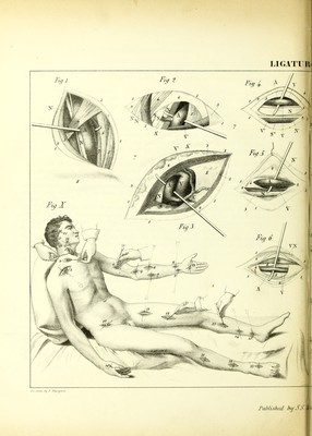
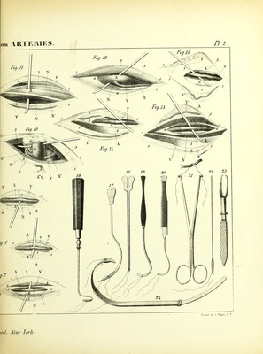
![Fig. 3. The Axillary Artery laid bare from hefore hackicards.—1, 1, 1, 1, Contour of the wound. 2, 2, The adipose and the aponeurotic layer. 3, 3, 3, Section of the pectoralis major divided in the direction of its fibres. 4, Upper border of the pectoralis minor—adipose pelotons and the deep-seated aponeurosis torn. A, The axillary artery brought forward by the sound, after having been disengaged from behind the vein, which is on the inside, and from the nervous branch nearest approximated to the chest, which is on the outside. A', A', The acromial and the anterior thoracic branch, which come by a common trunk from the principal artery. V, The axillary vein. V, The cephalic vein arriving from the arm in order to join the axillary, N, The first branch of the brachial plexus. Fig. 4. The Brachial Artery.—1, 1, 1, 1, Contour of the wound. 2, 2, The enveloping apo- neurosis. 3, 3, Another aponeurotic layer, which serves as a common sheath to the nervo- vascular fasciculus. 4, The fibrous partition which usually separates the artery from the median nerve. A, The brachial artery raised up from before backwards, that is, on its passage from the biceps muscle towards the inner portion of the triceps. V, V, Collateral veins. V, The basilic vein. N, The median nerve. N', The cutaneous nerve. N*, The ulnar (or cubital) nerve. Fig. 5. TJie Brachial Artery in the Fold or Bend of tlie Arm.—1, 1,1,1, Contour of the wound. 2, 2, 2, The aponeurosis divided and turned back to the outside. 3, A portion of the brachialis internus muscle. 4, Sheath of the artery. A, The artery raised up from without inwards, above the aponeurotic expansion of the biceps. V, Its collateral [i. e. associate or accompanv- ing] vein. N, The median nerve. N', A branch of the musculo-cutaneous nerve, separated aside by means of a hook, at the same time with the corresponding lip of the wound. Fig. 6. The Radial Artery above.—1, 1, 1, 1, The contour of the wound. 2, 2, First layer of the aponeurosis, divided and turned back on its borders. 3, The inner border of the supinator radii longus muscle. 4, Second layer of the aponeurosis. 5, The flexor digitorum sublimis [or superior or superficial] muscle. 6, The tendon of the pronator radii teres muscle. A, The artery raised up between the veins. V, Collateral veins. V', N, superficial vein and nerve. Fig. 7. The Ulnar Artery above.—1, 1, 1,1, Contour of the wound. 2, 2, 2, First aponeurotic layer. 3, The flexor digitorum sublimis. 4, The flexor carpi ulnaris muscle. 5, 5, The second aponeurotic layer. A, The artery disengaged from its envelopes and brought forward. V, The collateral veins situated on the radial side. N, The ulnar nerve. Fig. 8. Th£. Radial Artery beloiv.—1, 1, The contour of the wound. 2, 2, The aponeurosis.. 3, Tendon of the flexor carpi radialis muscle. 4, The pronator radii quadratus muscle behind the vessels. A, The artery separated from its veins, V. Fig. 9. The Ulnar Artery below.—1, 1, Contour of the wound. 2, 2, Aponeurosis. 3. The tendon of the flexor carpi ulnaris muscle pushed back to the inner side. 4, 4, The deep-seated layer of the aponeurosis. A, The artery, accompanied on its sides l)y the veins, V. N, Anterior branch of the ulnar nerve. Fig. 30. Ligature upon tlie Radial Artery between the Thumb and Forrfnger. THE LOWER EXTREMITY. Fig. 10. The External Iliac Artery.—1,1, 1, Contour of the wound. 2,2, Aponeurosis of the obliquus abdominis externus muscle. 3, 3, The obliquus abdominis internus muscle. 4, A](https://iiif.wellcomecollection.org/image/b21298610_0013.jp2/full/800%2C/0/default.jpg)


 No text description is available for this image
 No text description is available for this image
 No text description is available for this image