Volume 1
The household physician : a family guide to the preservation of health and to the domestic treatment of ailments and disease, with chapters on food and drugs and first aid in accidents and injuries / by J. McGregor-Robertson ; with an introduction by John G. McKendrick.
- M'Gregor-Robertson, J. (Joseph), 1858-1925
- Date:
- 1899
Licence: Public Domain Mark
Credit: The household physician : a family guide to the preservation of health and to the domestic treatment of ailments and disease, with chapters on food and drugs and first aid in accidents and injuries / by J. McGregor-Robertson ; with an introduction by John G. McKendrick. Source: Wellcome Collection.
54/602 page 18
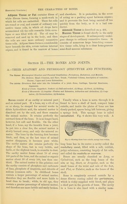
![THE STRUCTUKE OF BONE. [Sect. II. A. brane called endosteum, also rich iu blood- vessels. The bone itself seems so dry and hard as to have no moisture witliin it, nor any blood supply; but this is not the case, for blood- vessels pass into it from the periosteum through minute openings, and most long bones have be- sides a special artery (nutrient artery) enter- ing them to carry a due sui)ply of nourishment. If a very thin slice be taken from across a bone and examined under a microscope, magnifying about 300 diameters, an appearance is presented like that shown in Fig. 7, b. Little openings (a, b) are observed, and round them are ranged rings of bone, with little black bodies in them, from which fine dark lines branch out. These openings are canals cut across, called Haversian canals, after Havers, whofii-st described them; the black bodies are spaces, called lacunae (little lakes), in the bony plates, while the fine dark lines are very narrow canals which connect the lacunae with one another and with the Haver- sian canals. The narrow channels are called canaliculi. A, of the same figure, shows a section taken lengthways, by which the Haversian canals (a, b) have been opened up, not cut across, and are seen branching and communicat- ing with one another. Now, in these Haversian canals blood-vessels run; the lacunae contain little masses of a jelly-like mateidal—living protoplasm—processes from which pass along the canaliculi. c, of the figure, shows a lacuna (a), largely magnified, with its living mass of Early in the life of the child, months before it is born, there are no true bones in its body, their place being occupied by cartilage (gristle). The masses of cartilage have, however, the shape of bone, and it is out of them the bones are developed. Take such a one as the thigh- bone ; it is found that shortly before birth only the shaft has become true bone, the two ex- c? h Fig. 7.—Micro8copic.il appearance. A, of section of bone lengthwise, and 15, in cross section. C, A bone cell. protoplasm [h). Thus bones have not only a large blood supply obtained from the nutrient artery, and from the periosteum covering the outer, and the endosteum lining the iliner, sur- face, but contain innumerable little cells of living material, which are capable of drawing from the blood the sort of fof)d they require, and thus of maintaining a constant net-work of nourishing channels through the whole bone. tremities being still made of cartilage (Fig. 8, a). Just at birth a small deposit of earthy matter is found in the lower end (b. 2), which goes on extending. At one year of age a second deposit is seen in the upper end (c. 3). These little deposits, from which the bone forms, are called centres of ossification. The bone formation goes on from these centres till the ends are quite bony, and cartilage exists only at the place of junction between the shaft and the two extremities. The two ends are called epiphyses, and may readily be se])arated at this staire from the shaft. This is an accident which sometimes happens to children, and since it is at this point of junction that the jirincipal growth in the length of the bone occurs, such an accident may seriously interfere with the growth. The union between shaft and epiphyses does not take place till ma- turity, when further growth in the long direction ceases. Bones increase in thickness by growth from the inner surface of the periosteum. If, by ac- cident or disease, the periosteum be stripped off, no increase in thickness can take place, and the surface of the bone, deprived of its nourishment, will die; and> for a like reason, destruction of the inner lining membrane would impair the vitality of the inner surface. On the other hand the pei’iosteum may be kept in a higher state of activity than usual l)y constant irritation, as in chronic in- flammation. The result will be increased for- mation of new tissue and thickening of the bone. See Diseases of Bone.](https://iiif.wellcomecollection.org/image/b28124674_0001_0054.jp2/full/800%2C/0/default.jpg)
 No text description is available for this image
 No text description is available for this image
 No text description is available for this image