Volume 1
The science and art of surgery : a treatise on surgical injuries, diseases, and operations / by John Eric Erichsen.
- John Eric Erichsen
- Date:
- 1895
Licence: Public Domain Mark
Credit: The science and art of surgery : a treatise on surgical injuries, diseases, and operations / by John Eric Erichsen. Source: Wellcome Collection.
Provider: This material has been provided by The University of Leeds Library. The original may be consulted at The University of Leeds Library.
146/1274 (page 114)
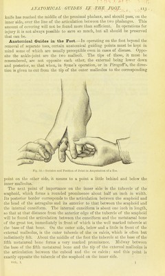
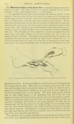
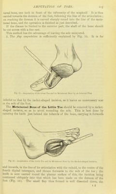
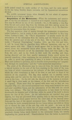
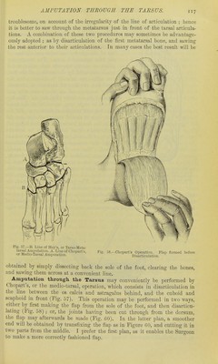
![The Metatarsal Bone of the Great Toe occasionally requires removal in whole or in part. The whole of the bone may be removed by one of two methods : 1, by a racket-shaped hicieioii ; 2, by the flap. ]. In Amputation by the racJcet-shaped imision the knife is entered on the dorsum of the foot about half an inch behind the tarsal end of the bone, and carried along the line of the bone to the level of the web between the first two toes ; it is then swept round the great toe, forming- an oval incision. On the outer side the knife must cut the skin at the point at which the web joins the great toe, and on the inner side the incision must correspond exactly to that on the outer. The oval part of the incisioii must not be commenced too soon ; in fact, it must be nearly circular, or the covering will be found insufficient (Pig. 54). The soft parts are then dissected as cleanly as possible from the upper, inner, and under surfaces of the bones, taking care not to leave the sesamoid bones in the flap. The knife is next passed between the head of the metatarsal bone and the next toe with its edge turned towards the toe. It will now be found that the head of the bone can be separated to some extent from that of the second metatarsal bone by forcing the forefinger of the left Fig. 54.—Amputation of the Great Toe and its Metatarsal Bone by the Racket-shaped Incision. hand between them. If it is found difficult to hold the bone with the fingers the lion forceps may be used. The knife is now passed to the outer side of the bone with its edge turned towards the tarsus, as in Fig. 54, while the meta- tarsal bone is forcibly pulled inwards so as to put the external ligaments of the tarso-metatarsal joint on the stretch. The knife being carried upwards with its edge closely applied to the metatarsal bone will divide the tense ligaments as soon as it touches them, and the joint will thus easily be opened Then, by twisting the bone while the knife is closely applied to its base, the removal is completed. If the knife be not closely ap])licd to the bone, and the ligaments put well on the stretch by forcibly pulling the toe inwards, it may enter the joint between the inner and middle cuneiform bones, and the latter bone may be removed. All danger of wounding the dorsal artery of the foot as it dips down between the two first metatarsal bones is avoided at the same time if the above directions are followed. 'J'he operation may be greatly facilitated by commencing the incision at the side of the foot immediately behind the tuberosity at the base of the meta-](https://iiif.wellcomecollection.org/image/b21510969_0001_0146.jp2/full/800%2C/0/default.jpg)