The experimental production of deafness in young animals by diet / by Edward Mellanby.
- Edward Mellanby
- Date:
- [1938?]
Licence: In copyright
Credit: The experimental production of deafness in young animals by diet / by Edward Mellanby. Source: Wellcome Collection.
27/32 (page 397)
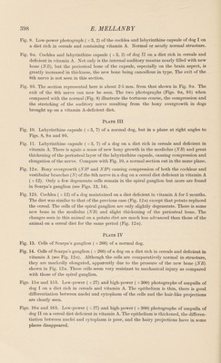
![Mellanby, E. [1930]. Brit. med. J. 1, 677. Mellanby, E. [1931]. Brain, 54, 247. Mellanby, E. [1933]. Edinb. med. J. 40, 197. Mellanby, E. [1934 a] J. Path. Bad. 38, 391. Mellanby, E. [19346]. Nutrition and Disease. Edinburgh and London: Oliver and Boyd. Mellanby, E. [1935]. Brain, 58, 141. Mellanby, E. [1937]. Chem. Ind. 56, 1054. Mellanby, E. [1938]. J. Physiol. 93, 42 P. Wittmaack, K. [1911]. Verh. dtsch. otol. Ges. 20, 295. Zimmerman, H. N. [1933]. J. exp. Med. 57, 215. EXPLANATION OF PLATES l-IV Plate I Fig. 1. Low-power photograph ( x 10) of cochlea of dog I. Basal diet with vitamin A added. On diet 10 months. It will be seen that the internal auditory meatus is clear and the cochlear branch of the 8th nerve (N) has an uninterrupted path to the brain. The spiral ganglion cells (Sp) are clearly seen (cp. Figs. 2, etc.). Fig. 2. Low-power photograph ( x 10) of cochlea of dog II. Basal diet as for dog I, but in this case no carotene or vitamin A was added. On diet 10 months. Two new bony masses (NB) practically filling the internal auditory meatus can be seen. The cells of the spiral ganglion (Sp') have disappeared and are replaced by connective tissue. Albuminoid coagula (G) can be well seen in scala tympani, indicating serous labyrin¬ thitis. Fig. 3. Middle whorl of cochlea ( x 27) of a normal dog showing organ of Corti and cells of spiral ganglion (Sp). Fig. 4. Middle whorl of cochlea ( x 27) of a dog on a diet containing much cereal and deficient in vitamin A. On diet 5 months. Many cells of the spiral ganglion (Sp') have disappeared together with the peripheral branches to the organ of Corti. Indications of serous labyrinthitis can be seen in the scala vestibuli. Plate II Fig. 5. Basal whorl of cochlea ( x 27) of dog II. Diet deficient in vitamin A. On diet 10 months. The cells of the spiral ganglion (Sp') have completely disappeared together with the peripheral nerve fibres to the organ of Corti. Albuminoid coagula (C) are obvious in both the scala tympani and scala vestibuli, indicating a condition of serous labyrinthitis. The organ of Corti is very abnormal. Fig. 6. High-power photograph ( x 266) of cells of the spiral ganglion of a normal dog. Note definite Nissl granules and well-defined nuclei. Fig. 7. High-power photograph ( x 266) of cells of the spiral ganglion of a dog whose diet was deficient in vitamin A. Nearly all the ganglion cells have completely degenerated and the few remaining cells are shrunken and the protoplasm is homogeneous. Cyto¬ plasm is devoid of granules.](https://iiif.wellcomecollection.org/image/b30631087_0027.jp2/full/800%2C/0/default.jpg)