The topographical anatomy of the head and neck of the horse / [O. Charnock Bradley].
- Orlando Charnock Bradley
- Date:
- 1923
Licence: In copyright
Credit: The topographical anatomy of the head and neck of the horse / [O. Charnock Bradley]. Source: Wellcome Collection.
108/248 page 92
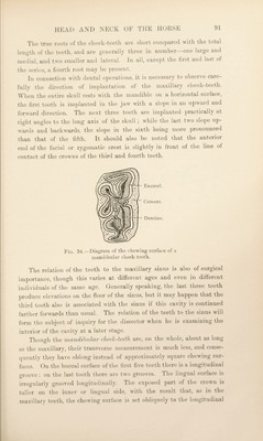
![plane of the tooth. Apart altogether from any structural arrangement, the maintenance of the slope on the chewing surfaces of the maxillary and mandibular teeth can be associated with the fact that the maxillary teeth of the two sides of the head are farther apart than are the two rows of mandibular teeth. The pattern assumed by the worn dental tissues on the mandi¬ bular teeth is different from and simpler than that of the maxillary teeth; for, while there are two infundibula, these are not closed on the lingual side until cement has been extensively developed. The conse¬ quence is that in the worn tooth the enamel fold lingual to each infundibulum is incomplete. Fig. 35.—The teeth after their embedded parts have ])een exposed by removal of the surrounding bone. With the exception of the last, which has usually three, each of the mandibular cheek-teeth has two relatively short roots. The first mandibular tooth is implanted in the jaw at right angles to the lower border of the mandible. The implantation of the rest of the teeth is oblique (downwards and backwards), the obliquity gradually increasing from the second to the last. The deciduous premolars are smaller than their representatives in the permanent dentition, their crowns are shorter, and a neck may be recognised. Dissection.—^The tongue, pharynx and larynx should now l)e removed, in order that an examination of the last-named may be proceeded with. In the first place, the thyro-hyoid and transverse hyoid muscles must be cleaned and defined. M.' THYREOHYOIDEUS.—The thyro-hyoid is a broad, flat muscle aris¬ ing from the body and thyroid process of the hyoid bone, and inserted](https://iiif.wellcomecollection.org/image/b29820066_0108.jp2/full/800%2C/0/default.jpg)