The topographical anatomy of the head and neck of the horse / [O. Charnock Bradley].
- Orlando Charnock Bradley
- Date:
- 1923
Licence: In copyright
Credit: The topographical anatomy of the head and neck of the horse / [O. Charnock Bradley]. Source: Wellcome Collection.
111/248 page 95
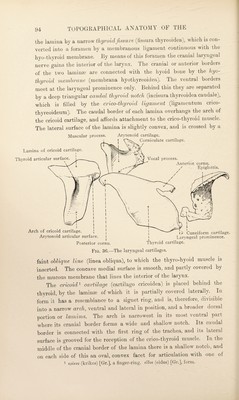
![the arytenoid cartilages (facies articularis arytcCnoidea). The caudal border of the lamina is thin and irregular, and overhangs the first ring of the trachea. The dorsal or outer surface of the lamina carries a median ridge-like miiscidav 'process (processus muscularis). On each side, at the junction of the arch and the lamina, there is a projecting, bracket-like articular surface for the reception of the caudal cornu of the thyroid (facies articularis thyreoidea). Tiie whole of the internal surface of the cricoid cartilage is smooth and covered by the laryngeal mucous membrane. The two arytenoid^ cartilages (cartilagines arytsenoideae), irregularly pyramidal in shape, rest on the cranial border of the lamina of the cricoid. The apex of each cartilage is directed towards the mouth, and is continued by a curved and sharply pointed corniculate cartilage (cartilage corniculata), which is yellow and elastic and pitted with depressions, while the arytenoid itself, like the thyroid and cricoid, is bluish in colour, rigid and smooth. The pointed tips of the corniculate cartilages curve upwards and backwards, and approach each other in the median plane, where, covered by mucous membrane, they form a characteristic projection in the undissected larynx. The triangular base of each arytenoid cartilage has a prominent ventral angle, the vocal process (processus vocalis) to which the vocal fold is attached, and a blunt lateral angle, the muscular jyrocess (processus muscularis), on which certain muscles terminate. The lateral part of the base carries an oval concave surface (facies articularis) for articulation with the cricoid cartilage. The lateral and dorsal surfaces of the arytenoid are concave, while the medial surface, larger than the other two, is flattened and covered by the laryngeal mucous membrane. The epiglottis ^ (cartilage epiglottica) has the form of a sessile leaf, resting within the laryngeal prominence of the thyroid by a broad base, from each corner of which an irregular rod of cartilage—the cuneiform^ cartilage (cartilage cuneiforme) projects towards the arytenoid. The anterior angle or apex of the epiglottis is free and sharply curved, pro¬ jects towards the base of the tongue, and rests on the edge of the soft palate. Of the two surfaces, the dorsal is entirely free and saddle- shaped (convex longitudinally and concave transversely), and is covered by firmly adherent mucous membrane. The ventral surface is also saddle-shaped (in the reverse direction) and partly free, but it affords attachment to the glosso-epiglottic mucous fold and the hyo-epiglottic muscle and ligament. The borders are thin and irregular. 1 apvTaiva (arytaiiia) [Gr.], a pitcher, a ladle, etoos (eidos) [Gr.], form. (epi) [Gr.], upon. yXuTTis (glottis) [Gr.], the mouth of the windpipe (Galen), the mouthpiece of a flute. 3 Cuneus [L.], a wedge.](https://iiif.wellcomecollection.org/image/b29820066_0111.jp2/full/800%2C/0/default.jpg)