The topographical anatomy of the head and neck of the horse / [O. Charnock Bradley].
- Orlando Charnock Bradley
- Date:
- 1923
Licence: In copyright
Credit: The topographical anatomy of the head and neck of the horse / [O. Charnock Bradley]. Source: Wellcome Collection.
113/248 page 97
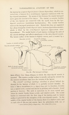
![the larynx is firmly adherent to the dorsal surface of the epiglottis, the medial surfaces of the arytenoid cartilages and over the vocal folds, where it is thin. Elsewhere in the vestibule and about the aditus it is thicker and loosely adherent to the subjacent structures. Dissection.—Remove the remains of the pharyngeal, sterno-thyroid, sterno-hyoid and thyro-hyoid muscle.s. Take care to ])reserve the laryngeal nerves and vessels. Now examine the structures on the ventral and lateral aspects of the larynx. The connection of the hyoid bone and the thyroid cartilage is established by the articulation of the extremities of the thyroid processes of the bone with the cranial cornua of the thyroid, and by the presence of the hyo-tkijroid membrane (membrana hyothyreoidea), which fills the gap between the hyoid bone and the thyroid cartilage, and is attached to the body and thyroid processes of the hyoid and to the anterior border of the thyroid laminaB. The joint between the bone and the thyroid cornu (articulatio hyothyreoidea) is a di arthrosis and is provided with a thick joint capsule. The movement in the joint is of the nature of a rotation about a transverse axis passing through the joints on the two sides of the larvnx. M. CRICOTHYREOTDEUS.—The ci'ico-thyroid muscle lies in the groove on the cricoid arch, from the lateral surface of which cartilage it takes its origin. Its insertion is into the caudal border and immediately adjacent part of the lateral surface of the thyroid lamina. By rotation of the cricoid cartilage about a transverse axis passing through the right and left cricothyroid joints, the muscle moves the anterior border of the cricoid lamina—and the bases of the arytenoid cartilages situated thereon—upwards and backwards, and thus tenses the vocal folds. The thin and membranous orico-thyvoid ligament (ligamentum cricothyreoideurn) is visible between the two crico-thyroid muscles, and fills the triangular interval between the thyroid and cricoid cartilages. The joint between the caudal thyroid cornu and the cricoid cartilage should be looked at now, but its complete examination may be deferred until the dissection of the larynx nears completion. The joint is a diarthrosis provided with the usual joint capsule attached round the articular margins of the two cartilages. Dissection.—Now turn to the dorsal aspect of the larynx. Remove the mucous membrane from the outer surface of the cricoid, arytenoid and corniculate cartilages, taking care not to destroy the cranial laryngeal and recurrent nerves. The origin of longitudinal muscular fibres of the msophagus from the interval between the cricoid and arytenoid cartilages 7](https://iiif.wellcomecollection.org/image/b29820066_0113.jp2/full/800%2C/0/default.jpg)