The topographical anatomy of the head and neck of the horse / [O. Charnock Bradley].
- Orlando Charnock Bradley
- Date:
- 1923
Licence: In copyright
Credit: The topographical anatomy of the head and neck of the horse / [O. Charnock Bradley]. Source: Wellcome Collection.
29/248 page 13
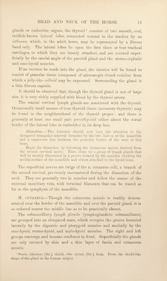
![distance parallel to tlie external maxillary vein on the surface of the internal pteryg’oid muscle, but soon the duct and vein come into contact: the duct being ventral to the vein, and the vein ventral to the external maxillary, artery. The three structures maintain this relative position as they turn round the margin of the mandible. The termination of the parotid duct will be examined in connection with the cheek. Lymph glands.' M. omohyoideus — Lig. cricotracheale. Parotid gland. P>ranch of 2nd cervical nerve. M. sternocephalicus.— M. mylohyoideus. M. digastricus. A. maxillaris interna. Mm. sternohyoideus. et omohyoideus. Parotid duct. --Hyoid bone. M. thyreohyoideus. Mem bran a hyothyreoidea. V. maxillaris externa. M. cricothyreoideus. M. sterno- thyreoideus. 1st tracheal cartilage. -M. omohyoideus. M. sternohyoideus. M. sternocephalicus. Fig. 4.—Dissection of the laryngeal region. First (left) and second (right) stages. M. DIGASTRICUS.—As its name indicates, the digastric^ muscle possesses two fleshy bellies joined by an intermediate tendon. At the present time it is possible to see the strong anterior belly only. This is inserted to the medial surface of the mandible close to the lower border of the molar part of this bone. M. MYLOGLOSSUS.—The pale, thin mylo-glossal - muscle consists of transverse fibres springing from the medial surface of the mandible, close to the alveolar border, from the symph3^sis to the third or fourth 1 dis (dis) [Hr.], double, yaar^p (ga.ster) [Gr.], belly. 2 fxv\r] (inyle) [Gr.], a mill ; referring to the cheek teeth. yXQaaa (glossa) [Gr.], the tongue.](https://iiif.wellcomecollection.org/image/b29820066_0029.jp2/full/800%2C/0/default.jpg)