The topographical anatomy of the head and neck of the horse / [O. Charnock Bradley].
- Orlando Charnock Bradley
- Date:
- 1923
Licence: In copyright
Credit: The topographical anatomy of the head and neck of the horse / [O. Charnock Bradley]. Source: Wellcome Collection.
34/248 page 18
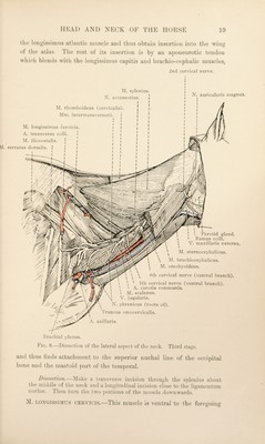
![vertebrge, the border of the wing of the atlas, and, by a thin aponeurotic tendon, to the mastoid portion of the temporal bone and the superior nuchal line of the occipital. The superficial face of the muscle is cleaned with some degree of difficulty because of the close adhesion to it of the cervical fascia and the cutaneous muscle; and its deep face is adherent to the omo-hyoid muscle. The thin temporal and occipital tendon is fused with the splenius and longissimus capitis, and is connected with the tendon of insertion of the sterno-cephalic muscle by an aponeurotic sheet. When the surface of the muscle has been divested of fascia, an imperfect division into two parts along a line indicated by the super¬ ficial rami of the ventral branches of the cervical nerves may be determined. The cleido-mastoid part (m. cleidomastoideus), attached to the temporal and occipital bones, partly overlaps the rest of the muscle (m. cleidotransversarius). Dissection.—The brachio-cephalic muscle should be cut across at the shoulder joint and turned aside. Its close adhesion to the underlying omo-hyoid muscle renders the reflection somewhat difficult. The superficial cervical lymph glands (lymphoglandula?. cervicales superficiales) occupy a triangular space bounded by the brachio-cephalic, omo-hyoid and deep pectoral muscles. They frequently reach and appear to blend with the caudal cervical glands. A. CERVICALIS ASCENDENS.—The ascending cervical ^ artery is one of the two divisions of the omo-cervical trunk. It crosses the lateral face of the jugular vein, is related for a short distance to the border of the prescapular part of the deep pectoral muscle and the superficial cervical lymph glands, and then runs between the omo-hyoid and brachio-cephalic muscles. M. SERRATUS VENTRALIS (CERVicis).—Though the ventral serrate ^ muscle belongs, strictly speaking, to the thoracic limb, the dissector of the neck should examine its attachment to the transverse processes of the last four (or five) cervical vertebra. This having been done, the remains of the muscle should be removed. M. SPLENIUS.—The thin triangular splenius ^ muscle arises by an aponeurotic tendon from the spinous processes of the second, third and fourth thoracic vertebrse, where it is confused with the origin of the dorsal serratus muscle, and from the ligamentum nuchse. It has fleshy insertions into the transverse processes of the fifth, fourth and third (sometimes second) cervical vertebrae. Some of its fibres blend with 1 Cervicalis [L.], pertaining to the neck {cervix). 2 Serratus (from serra, a saw) [L.], toothed or notched like the edge of a saw. 3 airXyjvLov (spleiiion) [Gr.], a bandage.](https://iiif.wellcomecollection.org/image/b29820066_0034.jp2/full/800%2C/0/default.jpg)