The topographical anatomy of the head and neck of the horse / [O. Charnock Bradley].
- Orlando Charnock Bradley
- Date:
- 1923
Licence: In copyright
Credit: The topographical anatomy of the head and neck of the horse / [O. Charnock Bradley]. Source: Wellcome Collection.
36/248 page 20
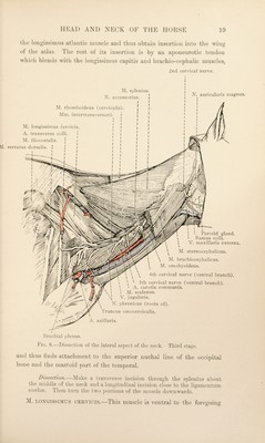
![—by which it is partly overlapped—and appears to be the direct continuation of the longrissimus dorsi. It arises from the transverse O processes of the first six or seven thoracic vertehrce, and ends on the transverse processes of the last four cervical vertebrm. M. vSCALENUS.—The brachial plexus of nerves divides the scalene ^ muscle of the horse into two parts. The dorsal and smaller portion of the muscle is attached to the cranial border of the first rib close to its vertebral end, and to the transverse process of the last cervical vertebra. The larger, ventral portion arises from the rough area on the cranial border and outer surface of the first rib proximal to a smooth groove produced by the subclavian vessels, and is inserted to the transverse processes of the sixth, fifth and fourth cervical vertebrae. N. CEUVICALIS SECUNDUS.—The present is a convenient time at which to examine the ventral branch of the second cervical nerve. The nerve appears at the lateral border of the caudal oblique muscle of the head. Underneath the brachio-cephalic muscle it communicates by anastomotic branches with the accessory nerve and with the ventral branches of the first and third cervical nerves, and is concerned in the formation of the nerve that has been noted as supplying the sterno- thyro-hyoid muscle. It then emerges between the two parts of the brachio-cephalic muscle and immediately divides into two branches. (1) The great auricidar nerve (n. auricularis magnus) follows the edge of the wing of the atlas and the contiguous border of the parotid gland, and ends in the skin covering the convex surface of the external ear. (2) The cutaneous cervical nerve (n. cutaneus colli) is connected with the ramus colli of the seventh cerebral nerve, and the combined nerve has already been found in association with the jugular vein. As has also been noted, a part of the cutaneous cervical nerve passes into the space between the two halves of the mandible. M. LONGISSIMUS CAPITIS. M. LONGISSIME^S ATLANTIS.—These two muscles are readily distinguished, but it is convenient to consider them together. They are long and narrow, extending the whole length of the neck, and lie medial to the splenius and longissimus cervicis. They arise in common from the transverse processes of the first two thoracic vertebrae by aponeurotic tendinous bands that are connected with the underlying semispinalis capitis muscle, and receive additional bundles of fibres from the articular processes of the last five (or six) cervical vertebrae. 1 Scalenus (scaleiios) [Gr.], uneven. The outline of the muscle is a triangle with unequal sides.](https://iiif.wellcomecollection.org/image/b29820066_0036.jp2/full/800%2C/0/default.jpg)