The topographical anatomy of the head and neck of the horse / [O. Charnock Bradley].
- Orlando Charnock Bradley
- Date:
- 1923
Licence: In copyright
Credit: The topographical anatomy of the head and neck of the horse / [O. Charnock Bradley]. Source: Wellcome Collection.
37/248 page 21
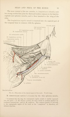
![\ V. jugularis. A. carotis communis. Ramus colli. V. maxillaris externa. M. bracliiocephalicus. Mm. intertransversarii. M. scalenus. N. phrenicus (roots of). 'fruncus omocervicalis. A. axillaris. Brachial plexus. Fi(t. 9.—Dissection of the lateivil aspect of the neck. Fourth stage. M. SEMISPIXALIS CAPITIS.^—(Jovered by the thin splenius muscle, ^ In animals like the dog, the seinis[)inalis capitis muscle is readily separated into two parts ;—(1) M. hiveattir cervicis, the more dorsal in position, marked by tendinous intersections ; and (2) M. complexus. Tlie common jn’actice of referring ^ to tlie semispinalis capitis of the horse as the “complexus” is, therefore, not without objection. J he more ventnil ol the two muscles, m. Ion;/ossiniii-s dilontls, ends in a stiong tendon that joins the flattened tendon common to the brachio¬ cephalic and splenius muscle.s, and is thus inserted to the wing of the atlas. J he Ionfjis,st/nils co/xtis vvascle is inserted into the mastoid part of the temporal bone in common with the splenius. M. longissimus capitis. M. longissimus atlantis. ' ! ! M. scmis])inalis capitis. i I I * I M. longissimus cervicis. | | I Ligamentum nuchic. A. transversa colli. I Parotid gland. 2nd cervical nerve. j M. splenius.](https://iiif.wellcomecollection.org/image/b29820066_0037.jp2/full/800%2C/0/default.jpg)